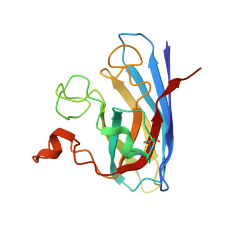
7T8H

G93A mutant of human SOD1 bound with MR6-26-2 in P21 space group


Experimental Data Snapshot

  • Method: X-RAY DIFFRACTION
  • Resolution: 1.50 Å
  • R-Value Free: 0.224 
  • R-Value Work: 0.186 

Starting Model: experimental
View more details

wwPDB Validation   3D Report Full Report


Ligand Structure Quality Assessment 


This is version 1.2 of the entry. See complete history


Literature

Ebselen analogues delay disease onset and its course in fALS by on-target SOD-1 engagement.

Watanabe, S.Amporndanai, K.Awais, R.Latham, C.Awais, M.O'Neill, P.M.Yamanaka, K.Hasnain, S.S.

(2024) Sci Rep 14: 12118-12118

  • DOI: https://doi.org/10.1038/s41598-024-62903-5
  • Primary Citation of Related Structures:  
    7T8E, 7T8F, 7T8G, 7T8H

  • PubMed Abstract: 

    Amyotrophic lateral sclerosis (ALS) selectively affects motor neurons. SOD1 is the first causative gene to be identified for ALS and accounts for at least 20% of the familial (fALS) and up to 4% of sporadic (sALS) cases globally with some geographical variability. The destabilisation of the SOD1 dimer is a key driving force in fALS and sALS. Protein aggregation resulting from the destabilised SOD1 is arrested by the clinical drug ebselen and its analogues (MR6-8-2 and MR6-26-2) by redeeming the stability of the SOD1 dimer. The in vitro target engagement of these compounds is demonstrated using the bimolecular fluorescence complementation assay with protein-ligand binding directly visualised by co-crystallography in G93A SOD1. MR6-26-2 offers neuroprotection slowing disease onset of SOD1 G93A mice by approximately 15 days. It also protected neuromuscular junction from muscle denervation in SOD1 G93A mice clearly indicating functional improvement.


  • Organizational Affiliation

    Department of Neuroscience and Pathobiology, Research Institute of Environmental Medicine, Nagoya University, Furo-Cho, Chikusa-Ku, Nagoya, 464-8601, Japan.


Macromolecules
Find similar proteins by:  (by identity cutoff)  |  3D Structure
Entity ID: 1
MoleculeChains Sequence LengthOrganismDetailsImage
Superoxide dismutase [Cu-Zn]A,
B [auth F]
153Homo sapiensMutation(s): 1 
Gene Names: SOD1
EC: 1.15.1.1
UniProt & NIH Common Fund Data Resources
Find proteins for P00441 (Homo sapiens)
Explore P00441 
Go to UniProtKB:  P00441
PHAROS:  P00441
GTEx:  ENSG00000142168 
Entity Groups  
Sequence Clusters30% Identity50% Identity70% Identity90% Identity95% Identity100% Identity
UniProt GroupP00441
Sequence Annotations
Expand
  • Reference Sequence
Small Molecules
Ligands 4 Unique
IDChains Name / Formula / InChI Key2D Diagram3D Interactions
Q8E (Subject of Investigation/LOI)
Query on Q8E

Download Ideal Coordinates CCD File 
E [auth A],
I [auth F]
~{N}-[(6-methoxypyridin-3-yl)methyl]-2-selanyl-benzamide
C14 H14 N2 O2 Se
SGEBTMJCWYBJSF-UHFFFAOYSA-N
SO4
Query on SO4

Download Ideal Coordinates CCD File 
G [auth A],
K [auth F]
SULFATE ION
O4 S
QAOWNCQODCNURD-UHFFFAOYSA-L
ZN
Query on ZN

Download Ideal Coordinates CCD File 
C [auth A],
F [auth A],
H [auth F],
J [auth F]
ZINC ION
Zn
PTFCDOFLOPIGGS-UHFFFAOYSA-N
ACT
Query on ACT

Download Ideal Coordinates CCD File 
D [auth A]ACETATE ION
C2 H3 O2
QTBSBXVTEAMEQO-UHFFFAOYSA-M
Experimental Data & Validation

Experimental Data

  • Method: X-RAY DIFFRACTION
  • Resolution: 1.50 Å
  • R-Value Free: 0.224 
  • R-Value Work: 0.186 
  • Space Group: P 1 21 1
Unit Cell:
Length ( Å )Angle ( ˚ )
a = 38.54α = 90
b = 68.478β = 105.024
c = 49.965γ = 90
Software Package:
Software NamePurpose
REFMACrefinement
iMOSFLMdata reduction
Aimlessdata scaling
MOLREPphasing

Structure Validation

View Full Validation Report


Ligand Structure Quality Assessment 


Entry History & Funding Information

Deposition Data


Funding OrganizationLocationGrant Number
Other privateUnited StatesWA1128

Revision History  (Full details and data files)

  • Version 1.0: 2023-01-25
    Type: Initial release
  • Version 1.1: 2023-10-25
    Changes: Data collection, Refinement description
  • Version 1.2: 2024-06-05
    Changes: Database references