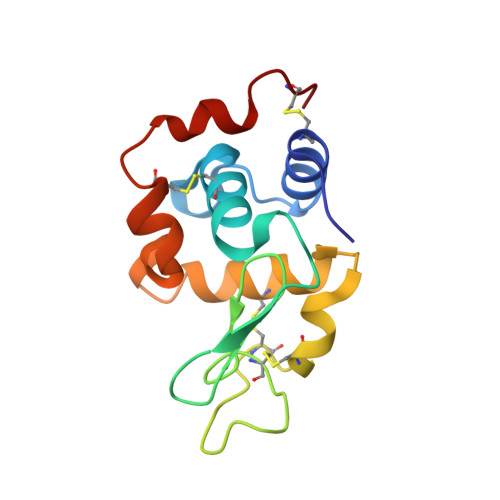
8A9D

Multicrystal room temperature structure of Lysozyme collected using a double multilayer monochromator.


Experimental Data Snapshot

  • Method: X-RAY DIFFRACTION
  • Resolution: 2.10 Å
  • R-Value Free: 0.249 
  • R-Value Work: 0.194 
  • R-Value Observed: 0.199 

Starting Model: experimental
View more details

wwPDB Validation   3D Report Full Report


This is version 1.5 of the entry. See complete history


Literature

Protein-to-structure pipeline for ambient-temperature in situ crystallography at VMXi.

Mikolajek, H.Sanchez-Weatherby, J.Sandy, J.Gildea, R.J.Campeotto, I.Cheruvara, H.Clarke, J.D.Foster, T.Fujii, S.Paulsen, I.T.Shah, B.S.Hough, M.A.

(2023) IUCrJ 10: 420-429

  • DOI: https://doi.org/10.1107/S2052252523003810
  • Primary Citation of Related Structures:  
    7ZCK, 8A9D, 8AR6, 8AR9, 8BRK, 8BRL, 8CIF

  • PubMed Abstract: 

    The utility of X-ray crystal structures determined under ambient-temperature conditions is becoming increasingly recognized. Such experiments can allow protein dynamics to be characterized and are particularly well suited to challenging protein targets that may form fragile crystals that are difficult to cryo-cool. Room-temperature data collection also enables time-resolved experiments. In contrast to the high-throughput highly automated pipelines for determination of structures at cryogenic temperatures widely available at synchrotron beamlines, room-temperature methodology is less mature. Here, the current status of the fully automated ambient-temperature beamline VMXi at Diamond Light Source is described, and a highly efficient pipeline from protein sample to final multi-crystal data analysis and structure determination is shown. The capability of the pipeline is illustrated using a range of user case studies representing different challenges, and from high and lower symmetry space groups and varied crystal sizes. It is also demonstrated that very rapid structure determination from crystals in situ within crystallization plates is now routine with minimal user intervention.


  • Organizational Affiliation

    Diamond Light Source Ltd, Harwell Science and Innovation Campus, Didcot OX11 0DE, United Kingdom.


Macromolecules
Find similar proteins by:  (by identity cutoff)  |  3D Structure
Entity ID: 1
MoleculeChains Sequence LengthOrganismDetailsImage
Lysozyme129Gallus gallusMutation(s): 0 
EC: 3.2.1.17
UniProt
Find proteins for P00698 (Gallus gallus)
Explore P00698 
Go to UniProtKB:  P00698
Entity Groups  
Sequence Clusters30% Identity50% Identity70% Identity90% Identity95% Identity100% Identity
UniProt GroupP00698
Sequence Annotations
Expand
  • Reference Sequence
Small Molecules
Ligands 1 Unique
IDChains Name / Formula / InChI Key2D Diagram3D Interactions
NA (Subject of Investigation/LOI)
Query on NA

Download Ideal Coordinates CCD File 
B [auth A]SODIUM ION
Na
FKNQFGJONOIPTF-UHFFFAOYSA-N
Experimental Data & Validation

Experimental Data

Unit Cell:
Length ( Å )Angle ( ˚ )
a = 78.485α = 90
b = 78.485β = 90
c = 38.139γ = 90
Software Package:
Software NamePurpose
PHENIXrefinement
PHENIXrefinement
xia2.multiplexdata reduction
xia2.multiplexdata scaling
PHASERphasing

Structure Validation

View Full Validation Report


Entry History & Funding Information

Deposition Data


Funding OrganizationLocationGrant Number
Not funded--

Revision History  (Full details and data files)

  • Version 1.0: 2022-07-20
    Type: Initial release
  • Version 1.1: 2023-05-17
    Changes: Database references
  • Version 1.2: 2023-05-31
    Changes: Database references
  • Version 1.3: 2023-07-19
    Changes: Data collection, Database references
  • Version 1.4: 2024-02-07
    Changes: Data collection, Refinement description
  • Version 1.5: 2024-11-06
    Changes: Structure summary