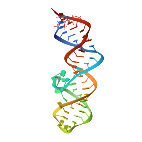
8XZQ

Crystal structure of folE riboswitch with 8-N Guanine


Experimental Data Snapshot

  • Method: X-RAY DIFFRACTION
  • Resolution: 1.83 Å
  • R-Value Free: 0.235 
  • R-Value Work: 0.209 
  • R-Value Observed: 0.210 

Starting Model: experimental
View more details

wwPDB Validation   3D Report Full Report


Ligand Structure Quality Assessment 


This is version 1.1 of the entry. See complete history


Literature

Structure-based characterization and compound identification of the wild-type THF class-II riboswitch.

Li, C.Xu, X.Geng, Z.Zheng, L.Song, Q.Shen, X.Wu, J.Zhao, J.Li, H.He, M.Tai, X.Zhang, L.Ma, J.Dong, Y.Ren, A.

(2024) Nucleic Acids Res 52: 8454-8465

  • DOI: https://doi.org/10.1093/nar/gkae377
  • Primary Citation of Related Structures:  
    8XZE, 8XZK, 8XZL, 8XZM, 8XZN, 8XZO, 8XZP, 8XZQ, 8XZR, 8XZW

  • PubMed Abstract: 

    Riboswitches are conserved regulatory RNA elements participating in various metabolic pathways. Recently, a novel RNA motif known as the folE RNA motif was discovered upstream of folE genes. It specifically senses tetrahydrofolate (THF) and is therefore termed THF-II riboswitch. To unravel the ligand recognition mechanism of this newly discovered riboswitch and decipher the underlying principles governing its tertiary folding, we determined both the free-form and bound-form THF-II riboswitch in the wild-type sequences. Combining structural information and isothermal titration calorimetry (ITC) binding assays on structure-based mutants, we successfully elucidated the significant long-range interactions governing the function of THF-II riboswitch and identified additional compounds, including alternative natural metabolites and potential lead compounds for drug discovery, that interact with THF-II riboswitch. Our structural research on the ligand recognition mechanism of the THF-II riboswitch not only paves the way for identification of compounds targeting riboswitches, but also facilitates the exploration of THF analogs in diverse biological contexts or for therapeutic applications.


  • Organizational Affiliation

    Life Sciences Institute, Second Affiliated Hospital of Zhejiang University School of Medicine, Zhejiang Key Laboratory of Biotherapy, Zhejiang University, Hangzhou 310058, China.


Macromolecules
Find similar nucleic acids by:  (by identity cutoff)  |  3D Structure
Entity ID: 1
MoleculeChains LengthOrganismImage
RNA (53-MER)53unidentified eubacterium clone A70
Sequence Annotations
Expand
  • Reference Sequence
Small Molecules
Ligands 3 Unique
IDChains Name / Formula / InChI Key2D Diagram3D Interactions
SPM
Query on SPM

Download Ideal Coordinates CCD File 
C [auth A]SPERMINE
C10 H26 N4
PFNFFQXMRSDOHW-UHFFFAOYSA-N
AZG (Subject of Investigation/LOI)
Query on AZG

Download Ideal Coordinates CCD File 
B [auth A]5-AMINO-1H-[1,2,3]TRIAZOLO[4,5-D]PYRIMIDIN-7-OL
C4 H4 N6 O
LPXQRXLUHJKZIE-UHFFFAOYSA-N
MG
Query on MG

Download Ideal Coordinates CCD File 
D [auth A]
E [auth A]
F [auth A]
G [auth A]
H [auth A]
D [auth A],
E [auth A],
F [auth A],
G [auth A],
H [auth A],
I [auth A],
J [auth A]
MAGNESIUM ION
Mg
JLVVSXFLKOJNIY-UHFFFAOYSA-N
Experimental Data & Validation

Experimental Data

  • Method: X-RAY DIFFRACTION
  • Resolution: 1.83 Å
  • R-Value Free: 0.235 
  • R-Value Work: 0.209 
  • R-Value Observed: 0.210 
  • Space Group: P 21 21 21
Unit Cell:
Length ( Å )Angle ( ˚ )
a = 49.268α = 90
b = 56.843β = 90
c = 62.778γ = 90
Software Package:
Software NamePurpose
PHENIXrefinement
HKL-3000data scaling
HKL-3000data reduction
PHASERphasing

Structure Validation

View Full Validation Report


Ligand Structure Quality Assessment 


Entry History & Funding Information

Deposition Data


Funding OrganizationLocationGrant Number
National Natural Science Foundation of China (NSFC)China--

Revision History  (Full details and data files)

  • Version 1.0: 2024-07-24
    Type: Initial release
  • Version 1.1: 2024-08-21
    Changes: Database references