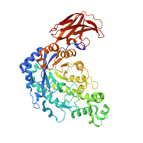
8ZRZ

The 1.26 angstrom resolution structure of Bacillus cereus beta-amylase in complex with maltose


Experimental Data Snapshot

  • Method: X-RAY DIFFRACTION
  • Resolution: 1.26 Å
  • R-Value Free: 0.165 
  • R-Value Work: 0.124 

Starting Model: experimental
View more details

wwPDB Validation   3D Report Full Report


Ligand Structure Quality Assessment 


This is version 1.2 of the entry. See complete history


Literature

Structural insight into sugar-binding modes of microbial ss-amylase.

Hirata, A.Mikami, B.

(2024) Biochem Biophys Res Commun 733: 150695-150695

  • DOI: https://doi.org/10.1016/j.bbrc.2024.150695
  • Primary Citation of Related Structures:  
    8ZRZ

  • PubMed Abstract: 

    ß-Amylase, which catalyses the release of ß-anomeric maltose from the non-reducing end of starch, is widely used in the food industry. Increasing its enzyme activity through protein engineering might improve the efficiency of food processing. To obtain detailed structural information to assist rationale design, here the crystal structure of Bacillus cereus β-amylase (BCB) complexed with maltose was determined by molecular replacement and refined using anisotropic temperature factors to 1.26 Å resolution with R work /R free factors of 12.4/15.7 %. The structure contains six maltose and one glucose molecules, of which two maltose and one glucose are bound at sites not previously observed in BCB structures. These three new sugar-binding sites are located on the surface and likely to be important in enhancing the degradation of raw-starch granules. In the active site of BCB, two maltose molecules are bound in tandem at subsites -2 ∼ -1 and +1 ∼ +2. Notably, the conformation of the glucose moiety bound at subsite -1 is a mixture of α-anomeric distorted 1,4 B boat and 4 C 1 chair forms, while those at subsites -2, +1 ∼ +2 are all in the 4 C 1 chair forms. The O1 of the distorted α-glucose residue at subsite -1 occupies the position of the putative catalytic water, forming a hydrogen bond with OE1 of Glu367 (base catalyst), suggesting that this distorted sugar is not involved in catalysis. Together, these findings pave the way for further improving the functionality of microbial ß-amylase enzymes.


  • Organizational Affiliation

    Department of Natural Science, Graduate School of Technology, Industrial and Social Science, Tokushima University, 2-1 Minamijosanjimacho, Tokushima, Tokushima, 770-8506, Japan. Electronic address: ahirata@tokushima-u.ac.jp.


Macromolecules
Find similar proteins by:  (by identity cutoff)  |  3D Structure
Entity ID: 1
MoleculeChains Sequence LengthOrganismDetailsImage
Beta-amylase516Bacillus cereusMutation(s): 0 
Gene Names: spoII
EC: 3.2.1.2
UniProt
Find proteins for P36924 (Bacillus cereus)
Explore P36924 
Go to UniProtKB:  P36924
Entity Groups  
Sequence Clusters30% Identity50% Identity70% Identity90% Identity95% Identity100% Identity
UniProt GroupP36924
Sequence Annotations
Expand
  • Reference Sequence
Oligosaccharides

Help

Entity ID: 2
MoleculeChains Length2D Diagram Glycosylation3D Interactions
alpha-D-glucopyranose-(1-4)-alpha-D-glucopyranoseB [auth C],
C [auth F],
E [auth D]
2N/A
Glycosylation Resources
GlyTouCan:  G07411ON
GlyCosmos:  G07411ON
Entity ID: 3
MoleculeChains Length2D Diagram Glycosylation3D Interactions
alpha-D-glucopyranose-(1-4)-beta-D-glucopyranoseD [auth E]2N/A
Glycosylation Resources
GlyTouCan:  G66120GK
GlyCosmos:  G66120GK
Entity ID: 4
MoleculeChains Length2D Diagram Glycosylation3D Interactions
alpha-D-glucopyranose-(1-4)-alpha-D-glucopyranose-(1-4)-alpha-D-glucopyranose-(1-4)-alpha-D-glucopyranoseF [auth B]4N/A
Glycosylation Resources
GlyTouCan:  G87171PZ
GlyCosmos:  G87171PZ
GlyGen:  G87171PZ
Small Molecules
Ligands 4 Unique
IDChains Name / Formula / InChI Key2D Diagram3D Interactions
BGC
Query on BGC

Download Ideal Coordinates CCD File 
Q [auth A]beta-D-glucopyranose
C6 H12 O6
WQZGKKKJIJFFOK-VFUOTHLCSA-N
SO4
Query on SO4

Download Ideal Coordinates CCD File 
H [auth A]SULFATE ION
O4 S
QAOWNCQODCNURD-UHFFFAOYSA-L
GOL
Query on GOL

Download Ideal Coordinates CCD File 
I [auth A]
J [auth A]
K [auth A]
L [auth A]
M [auth A]
I [auth A],
J [auth A],
K [auth A],
L [auth A],
M [auth A],
N [auth A],
O [auth A],
P [auth A]
GLYCEROL
C3 H8 O3
PEDCQBHIVMGVHV-UHFFFAOYSA-N
CA
Query on CA

Download Ideal Coordinates CCD File 
G [auth A]CALCIUM ION
Ca
BHPQYMZQTOCNFJ-UHFFFAOYSA-N
Experimental Data & Validation

Experimental Data

  • Method: X-RAY DIFFRACTION
  • Resolution: 1.26 Å
  • R-Value Free: 0.165 
  • R-Value Work: 0.124 
  • Space Group: P 1 21 1
Unit Cell:
Length ( Å )Angle ( ˚ )
a = 56.974α = 90
b = 89.843β = 103.09
c = 66.035γ = 90
Software Package:
Software NamePurpose
PHENIXphasing
SHELXL-97refinement
HKL-2000data reduction
HKL-2000data scaling

Structure Validation

View Full Validation Report


Ligand Structure Quality Assessment 


Entry History & Funding Information

Deposition Data


Funding OrganizationLocationGrant Number
Japan Society for the Promotion of Science (JSPS)Japan22K05441

Revision History  (Full details and data files)

  • Version 1.0: 2024-09-25
    Type: Initial release
  • Version 1.1: 2024-10-02
    Changes: Database references
  • Version 1.2: 2024-10-23
    Changes: Structure summary