1B54 | pdb_00001b54

CRYSTAL STRUCTURE OF A YEAST HYPOTHETICAL PROTEIN-A STRUCTURE FROM BNL'S HUMAN PROTEOME PROJECT


Experimental Data Snapshot

  • Method: X-RAY DIFFRACTION
  • Resolution: 2.10 Å
  • R-Value Free: 
    0.303 (Depositor), 0.270 (DCC) 
  • R-Value Work: 
    0.235 (Depositor), 0.220 (DCC) 
  • R-Value Observed: 
    0.235 (Depositor) 

wwPDB Validation   3D Report Full Report


Ligand Structure Quality Assessment 

Created with Raphaël 2.3.0Worse 01 BetterLigand structure goodness of fit to experimental dataBest fitted PLPClick on this verticalbar to view details

This is version 1.4 of the entry. See complete history


Literature

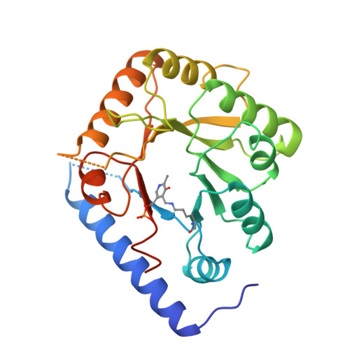
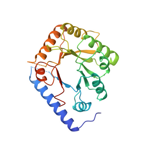
Structure of a yeast hypothetical protein selected by a structural genomics approach.

Eswaramoorthy, S.Gerchman, S.Graziano, V.Kycia, H.Studier, F.W.Swaminathan, S.

(2003) Acta Crystallogr D Biol Crystallogr 59: 127-135

  • DOI: https://doi.org/10.1107/s0907444902018012
  • Primary Citation of Related Structures:  
    1B54, 1CT5

  • PubMed Abstract: 

    Yeast hypothetical protein YBL036C (SWISS-PROT P38197), initially thought to be a member of an 11-protein family, was selected for crystal structure determination since no structural or functional information was available. The structure has been determined independently by MIR and MAD methods to 2.0 A resolution. The MAD structure was determined largely through automated model building. The protein folds as a TIM barrel beginning with a long N-terminal helix, in contrast to the classic triose phosphate isomerase (TIM) structure, which begins with a beta-strand. A cofactor, pyridoxal 5'-phosphate, is covalently bound near the C-terminal end of the barrel, the usual active site in TIM-barrel folds. A single-domain monomeric molecule, this yeast protein resembles the N-terminal domain of alanine racemase or ornithine decarboxylase, both of which are two-domain dimeric proteins. The yeast protein has been shown to have amino-acid racemase activity. Although selected as a member of a protein family having no obvious relationship to proteins of known structure, the protein fold turned out to be a well known and widely distributed fold. This points to the need for a more comprehensive base of structural information and better structure-modeling tools before the goal of structure prediction from amino-acid sequences can be realised. In this case, similarity to a known structure allowed inferences to be made about the structure and function of a widely distributed protein family.


  • Organizational Affiliation

    Biology Department, Brookhaven National Laboratory, Upton, NY 11973, USA.


Macromolecules
Find similar proteins by:  (by identity cutoff)  |  3D Structure
Entity ID: 1
MoleculeChains Sequence LengthOrganismDetailsImage
YEAST HYPOTHETICAL PROTEIN257Saccharomyces cerevisiaeMutation(s): 0 
Gene Names: SGD: YBL 036C
UniProt
Find proteins for P38197 (Saccharomyces cerevisiae (strain ATCC 204508 / S288c))
Explore P38197 
Go to UniProtKB:  P38197
Entity Groups  
Sequence Clusters30% Identity50% Identity70% Identity90% Identity95% Identity100% Identity
UniProt GroupP38197
Sequence Annotations
Expand
  • Reference Sequence
Small Molecules
Ligands 1 Unique
IDChains Name / Formula / InChI Key2D Diagram3D Interactions
PLP
Query on PLP

Download Ideal Coordinates CCD File 
B [auth A]PYRIDOXAL-5'-PHOSPHATE
C8 H10 N O6 P
NGVDGCNFYWLIFO-UHFFFAOYSA-N
Experimental Data & Validation

Experimental Data

  • Method: X-RAY DIFFRACTION
  • Resolution: 2.10 Å
  • R-Value Free:  0.303 (Depositor), 0.270 (DCC) 
  • R-Value Work:  0.235 (Depositor), 0.220 (DCC) 
  • R-Value Observed: 0.235 (Depositor) 
Space Group: P 21 21 21
Unit Cell:
Length ( Å )Angle ( ˚ )
a = 48.58α = 90
b = 65.71β = 90
c = 91.94γ = 90
Software Package:
Software NamePurpose
PHASESphasing
X-PLORmodel building
X-PLORrefinement
DENZOdata reduction
SCALEPACKdata scaling
X-PLORphasing

Structure Validation

View Full Validation Report


Ligand Structure Quality Assessment 

Created with Raphaël 2.3.0Worse 01 BetterLigand structure goodness of fit to experimental dataBest fitted PLPClick on this verticalbar to view details

Entry History 

Deposition Data

Revision History  (Full details and data files)

  • Version 1.0: 1999-01-27
    Type: Initial release
  • Version 1.1: 2008-03-03
    Changes: Version format compliance
  • Version 1.2: 2011-07-13
    Changes: Non-polymer description, Version format compliance
  • Version 1.3: 2021-02-03
    Changes: Derived calculations, Structure summary
  • Version 1.4: 2025-03-26
    Changes: Data collection, Database references, Structure summary