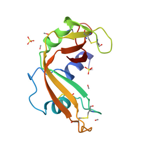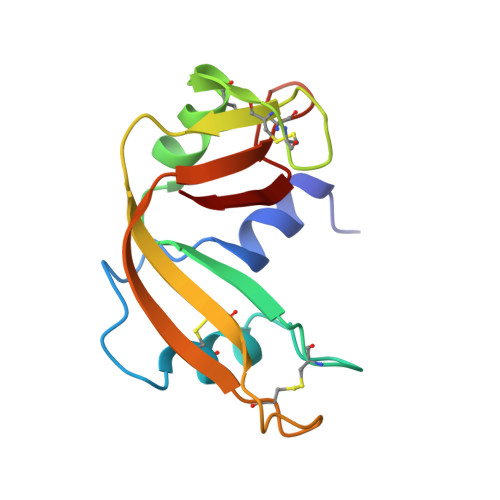
Structure of crystal form IX of bovine pancreatic ribonuclease A.
Dung, M.H., Bell, J.A.(1997) Acta Crystallogr D Biol Crystallogr 53: 419-425
- PubMed: 15299907
- DOI: https://doi.org/10.1107/S0907444997000929
- Primary Citation of Related Structures:
1BEL - PubMed Abstract:
The X-ray structure of crystal form IX of bovine pancre- atic ribonuclease A (space group P2(1)2(1)2(1)) is reported at 1.6 A resolution. The structure was refined to an R factor of 15.0% and includes coordinates for two sulfate ions, four methanol molecules and 82 waters. The structure could be superimposed on the highest resolution crystal structure of bovine pancreatic fibonuclease available (in space group P2(1)) with an r.m.s, difference in main-chain atomic positions of 0.51 A. Most of the larger differences between the two structures could be related to crystal lattice contacts. Superposition of the new structure with eight other structures of ribonuclease in six crystal forms resulted in an r.m.s, deviation from the average structure of 0.43 A for all main-chain atoms. This similarity among structures exists in spite of the fact that all nine molecules are in different crystal environments.
Organizational Affiliation:
Department of Chemistry and Center for Biophysics, Rensselaer Polytechnic Institute, Troy, NY 12180, USA.