2YQ5 | pdb_00002yq5

Crystal Structure of D-isomer specific 2-hydroxyacid dehydrogenase from Lactobacillus delbrueckii ssp. bulgaricus: NAD complexed form


Experimental Data Snapshot

  • Method: X-RAY DIFFRACTION
  • Resolution: 2.75 Å
  • R-Value Free: 
    0.259 (Depositor), 0.260 (DCC) 
  • R-Value Work: 
    0.188 (Depositor), 0.190 (DCC) 
  • R-Value Observed: 
    0.191 (Depositor) 

Starting Model: experimental
View more details

wwPDB Validation   3D Report Full Report


Ligand Structure Quality Assessment 

Created with Raphaël 2.3.0Worse 01 BetterLigand structure goodness of fit to experimental dataBest fitted NADClick on this verticalbar to view details

This is version 1.2 of the entry. See complete history


Literature

Structural Characterization of D-Isomer Specific 2-Hydroxyacid Dehydrogenase from Lactobacillus Delbrueckii Ssp. Bulgaricus

Holton, S.J.Anandhakrishnan, M.Geerlof, A.Wilmanns, M.

(2013) J Struct Biol 181: 179

  • DOI: https://doi.org/10.1016/j.jsb.2012.10.009
  • Primary Citation of Related Structures:  
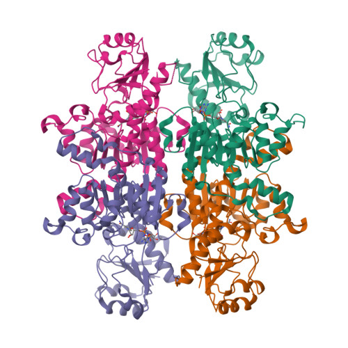
    2YQ4, 2YQ5

  • PubMed Abstract: 

    Hydroxyacid dehydrogenases, responsible for the stereospecific conversion of 2-keto acids to 2-hydroxyacids in lactic acid producing bacteria, have a range of biotechnology applications including antibiotic synthesis, flavor development in dairy products and the production of valuable synthons. The genome of Lactobacillus delbrueckii ssp. bulgaricus, a member of the heterogeneous group of lactic acid bacteria, encodes multiple hydroxyacid dehydrogenases whose structural and functional properties remain poorly characterized. Here, we report the apo and coenzyme NAD⁺ complexed crystal structures of the L. bulgaricusD-isomer specific 2-hydroxyacid dehydrogenase, D2-HDH. Comparison with closely related members of the NAD-dependent dehydrogenase family reveals that whilst the D2-HDH core fold is structurally conserved, the substrate-binding site has a number of non-canonical features that may influence substrate selection and thus dictate the physiological function of the enzyme.


  • Organizational Affiliation

    EMBL c/o DESY, Notkestrasse 85, D-22603 Hamburg, Germany. sjholton@googlemail.com


Macromolecules
Find similar proteins by:  (by identity cutoff)  |  3D Structure
Entity ID: 1
MoleculeChains Sequence LengthOrganismDetailsImage
D-ISOMER SPECIFIC 2-HYDROXYACID DEHYDROGENASE
A, B, C, D
343Lactobacillus delbrueckii subsp. bulgaricusMutation(s): 0 
UniProt
Find proteins for Q1GAA2 (Lactobacillus delbrueckii subsp. bulgaricus (strain ATCC 11842 / DSM 20081 / BCRC 10696 / JCM 1002 / NBRC 13953 / NCIMB 11778 / NCTC 12712 / WDCM 00102 / Lb 14))
Explore Q1GAA2 
Go to UniProtKB:  Q1GAA2
Entity Groups  
Sequence Clusters30% Identity50% Identity70% Identity90% Identity95% Identity100% Identity
UniProt GroupQ1GAA2
Sequence Annotations
Expand
  • Reference Sequence
Experimental Data & Validation

Experimental Data

  • Method: X-RAY DIFFRACTION
  • Resolution: 2.75 Å
  • R-Value Free:  0.259 (Depositor), 0.260 (DCC) 
  • R-Value Work:  0.188 (Depositor), 0.190 (DCC) 
  • R-Value Observed: 0.191 (Depositor) 
Space Group: P 21 21 21
Unit Cell:
Length ( Å )Angle ( ˚ )
a = 92.718α = 90
b = 110.012β = 90
c = 147.736γ = 90
Software Package:
Software NamePurpose
REFMACrefinement
XDSdata reduction
SCALAdata scaling
PHASERphasing

Structure Validation

View Full Validation Report


Ligand Structure Quality Assessment 

Created with Raphaël 2.3.0Worse 01 BetterLigand structure goodness of fit to experimental dataBest fitted NADClick on this verticalbar to view details

Entry History 

Deposition Data

Revision History  (Full details and data files)

  • Version 1.0: 2012-11-21
    Type: Initial release
  • Version 1.1: 2013-02-20
    Changes: Database references
  • Version 1.2: 2023-12-20
    Changes: Data collection, Database references, Derived calculations, Other, Refinement description