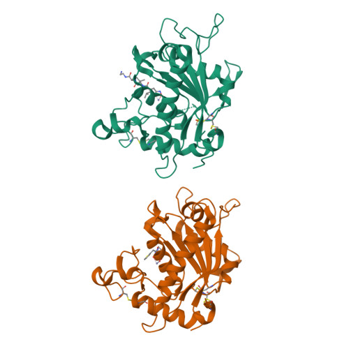
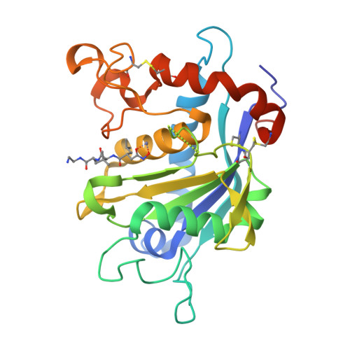
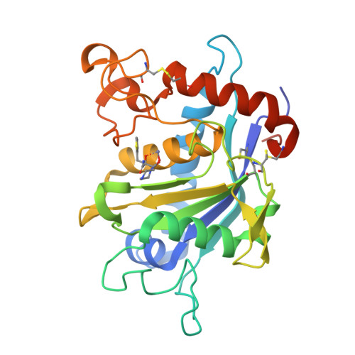
3KMC | pdb_00003kmc

Crystal structure of catalytic domain of TACE with tartrate-based inhibitor


Experimental Data Snapshot

  • Method: X-RAY DIFFRACTION
  • Resolution: 1.80 Å
  • R-Value Free: 
    0.259 (Depositor), 0.260 (DCC) 
  • R-Value Work: 
    0.224 (Depositor), 0.220 (DCC) 

Starting Model: experimental
View more details

wwPDB Validation   3D Report Full Report


Ligand Structure Quality Assessment 

Created with Raphaël 2.3.0Worse 01 BetterLigand structure goodness of fit to experimental dataBest fitted 403Click on this verticalbar to view detailsBest fitted INNClick on this verticalbar to view details

This is version 1.6 of the entry. See complete history


Literature

The discovery of novel tartrate-based TNF-alpha converting enzyme (TACE) inhibitors.

Rosner, K.E.Guo, Z.Orth, P.Shipps, G.W.Belanger, D.B.Chan, T.Y.Curran, P.J.Dai, C.Deng, Y.Girijavallabhan, V.M.Hong, L.Lavey, B.J.Lee, J.F.Li, D.Liu, Z.Popovici-Muller, J.Ting, P.C.Vaccaro, H.Wang, L.Wang, T.Yu, W.Zhou, G.Niu, X.Sun, J.Kozlowski, J.A.Lundell, D.J.Madison, V.McKittrick, B.Piwinski, J.J.Shih, N.Y.Arshad Siddiqui, M.Strickland, C.O.

(2009) Bioorg Med Chem Lett 20: 1189-1193

  • DOI: https://doi.org/10.1016/j.bmcl.2009.12.004
  • Primary Citation of Related Structures:  
    3KMC, 3KME

  • PubMed Abstract: 

    A novel series of TNF-alpha convertase (TACE) inhibitors which are non-hydroxamate have been discovered. These compounds are bis-amides of L-tartaric acid (tartrate) and coordinate to the active site zinc in a tridentate manner. They are selective for TACE over other MMP's. We report the first X-ray crystal structure for a tartrate-based TACE inhibitor.


  • Organizational Affiliation

    Department of Medicinal Chemistry, Schering-Plough Research Institute Cambridge, 320 Bent Street, Cambridge, MA 02141, United States.


Macromolecules
Find similar proteins by:  (by identity cutoff)  |  3D Structure
Entity ID: 1
MoleculeChains Sequence LengthOrganismDetailsImage
TNF-alpha-converting enzyme
A, B
270Homo sapiensMutation(s): 3 
Gene Names: ADAM17CSVPTACE
EC: 3.4.24.86
UniProt & NIH Common Fund Data Resources
Find proteins for P78536 (Homo sapiens)
Explore P78536 
Go to UniProtKB:  P78536
PHAROS:  P78536
GTEx:  ENSG00000151694 
Entity Groups  
Sequence Clusters30% Identity50% Identity70% Identity90% Identity95% Identity100% Identity
UniProt GroupP78536
Sequence Annotations
Expand
  • Reference Sequence
Small Molecules
Binding Affinity Annotations 
IDSourceBinding Affinity
403 BindingDB:  3KMC Ki: 400 (nM) from 1 assay(s)
Experimental Data & Validation

Experimental Data

  • Method: X-RAY DIFFRACTION
  • Resolution: 1.80 Å
  • R-Value Free:  0.259 (Depositor), 0.260 (DCC) 
  • R-Value Work:  0.224 (Depositor), 0.220 (DCC) 
Space Group: P 21 21 21
Unit Cell:
Length ( Å )Angle ( ˚ )
a = 74.313α = 90
b = 76.103β = 90
c = 103.768γ = 90
Software Package:
Software NamePurpose
CrystalCleardata collection
BUSTERrefinement
HKL-2000data reduction
HKL-2000data scaling

Structure Validation

View Full Validation Report


Ligand Structure Quality Assessment 

Created with Raphaël 2.3.0Worse 01 BetterLigand structure goodness of fit to experimental dataBest fitted 403Click on this verticalbar to view detailsBest fitted INNClick on this verticalbar to view details

Entry History 

Deposition Data

  • Released Date: 2009-12-22 
  • Deposition Author(s): Orth, P.

Revision History  (Full details and data files)

  • Version 1.0: 2009-12-22
    Type: Initial release
  • Version 1.1: 2011-07-13
    Changes: Non-polymer description, Version format compliance
  • Version 1.2: 2013-02-27
    Changes: Other
  • Version 1.3: 2017-11-01
    Changes: Refinement description
  • Version 1.4: 2021-10-13
    Changes: Database references, Derived calculations
  • Version 1.5: 2023-09-06
    Changes: Data collection, Refinement description
  • Version 1.6: 2024-11-27
    Changes: Structure summary