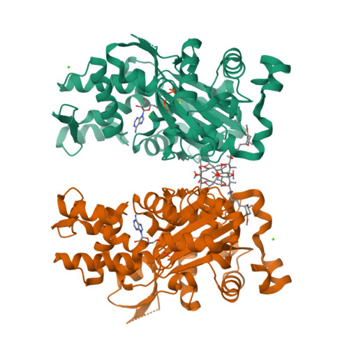
Two molecules of lobophorolide cooperate to stabilize an actin dimer using both their "ring" and "tail" region.
Blain, J.C., Mok, Y.F., Kubanek, J., Allingham, J.S.(2010) Chem Biol 17: 802-807
- PubMed: 20797609
- DOI: https://doi.org/10.1016/j.chembiol.2010.06.010
- Primary Citation of Related Structures:
3M6G - PubMed Abstract:
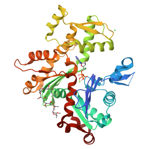
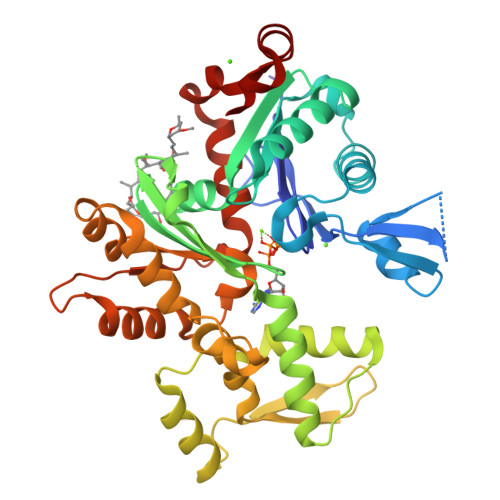
Actin filament-disrupting marine macrolides are promising templates from which to design therapeutics against cancer and other diseases that co-opt the actin cytoskeleton. Typically, these macrolides form either a 1:1 or 2:1 actin-macrolide complex where their aliphatic side chain, or "tail," has been reported to convey the major determinant of cytotoxicity. We now report the structure of the marine macrolide lobophorolide bound to actin with a unique 2:2 stoichiometry in which two lobophorolide molecules cooperate to form a dimerization interface that is composed entirely of the macrolide "ring" region, and each molecule of lobophorolide interacts with both actin subunits via their ring and tail regions to tether the subunits together. This binding mode imposes multiple barriers against microfilament stability and holds important implications for development of actin-targeting drugs and the evolution of macrolide biosynthetic enzymes.
Organizational Affiliation:
Department of Biochemistry, Queen's University, Kingston, ON K7L 3N6, Canada.