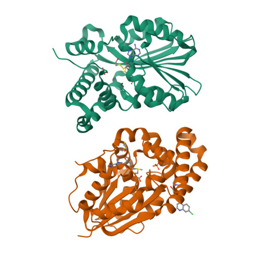
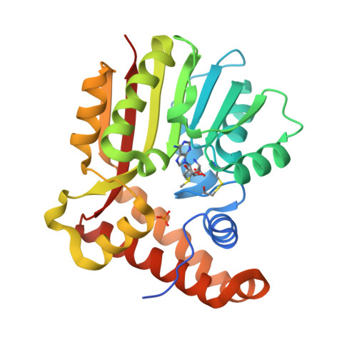
Phosphoethanolamine N-methyl transferase is a Malarial drug target
Lukk, T., Nair, S.K., Mamoun, C.B.To be published.
Experimental Data Snapshot
Starting Model: experimental
View more details
Entity ID: 1 | |||||
|---|---|---|---|---|---|
| Molecule | Chains | Sequence Length | Organism | Details | Image |
| Phosphoethanolamine N-methyltransferase, putative | 267 | Plasmodium vivax Sal-1 | Mutation(s): 0 Gene Names: PMT, PVX_083045 EC: 2.1.1.103 | 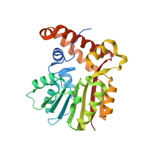 | |
UniProt | |||||
Find proteins for A5K867 (Plasmodium vivax (strain Salvador I)) Explore A5K867 Go to UniProtKB: A5K867 | |||||
Entity Groups | |||||
| Sequence Clusters | 30% Identity50% Identity70% Identity90% Identity95% Identity100% Identity | ||||
| UniProt Group | A5K867 | ||||
Sequence AnnotationsExpand | |||||
| |||||
| Ligands 4 Unique | |||||
|---|---|---|---|---|---|
| ID | Chains | Name / Formula / InChI Key | 2D Diagram | 3D Interactions | |
| SAM Query on SAM | C [auth A], F [auth B] | S-ADENOSYLMETHIONINE C15 H22 N6 O5 S MEFKEPWMEQBLKI-FCKMPRQPSA-N |  | ||
| CQA Query on CQA | H [auth B] | 4-[(7-CHLOROQUINOLIN-4-YL)AMINO]-2-[(DIETHYLAMINO)METHYL]PHENOL C20 H22 Cl N3 O OVCDSSHSILBFBN-UHFFFAOYSA-N |  | ||
| PO4 Query on PO4 | D [auth A], G [auth B] | PHOSPHATE ION O4 P NBIIXXVUZAFLBC-UHFFFAOYSA-K |  | ||
| BME Query on BME | E [auth A], I [auth B] | BETA-MERCAPTOETHANOL C2 H6 O S DGVVWUTYPXICAM-UHFFFAOYSA-N |  | ||
| Length ( Å ) | Angle ( ˚ ) |
|---|---|
| a = 37.44 | α = 90 |
| b = 86.62 | β = 103.23 |
| c = 78.27 | γ = 90 |
| Software Name | Purpose |
|---|---|
| PHASER | phasing |
| PHENIX | refinement |
| PDB_EXTRACT | data extraction |
| HKL-2000 | data collection |
| XDS | data reduction |
| XSCALE | data scaling |