4QX6 | pdb_00004qx6

CRYSTAL STRUCTURE OF GLYCERALDEHYDE-3-PHOSPHATE DEHYDROGENASE FROM STREPTOCOCCUS AGALACTIAE NEM316 at 2.46 ANGSTROM RESOLUTION


Experimental Data Snapshot

  • Method: X-RAY DIFFRACTION
  • Resolution: 2.46 Å
  • R-Value Free: 
    0.238 (Depositor), 0.240 (DCC) 
  • R-Value Work: 
    0.204 (Depositor), 0.210 (DCC) 
  • R-Value Observed: 
    0.206 (Depositor) 

Starting Model: experimental
View more details

wwPDB Validation   3D Report Full Report


Ligand Structure Quality Assessment 

Created with Raphaël 2.3.0Worse 01 BetterLigand structure goodness of fit to experimental dataBest fitted NADClick on this verticalbar to view details

This is version 1.4 of the entry. See complete history


Literature

Structure of Streptococcus agalactiae glyceraldehyde-3-phosphate dehydrogenase holoenzyme reveals a novel surface.

Ayres, C.A.Schormann, N.Senkovich, O.Fry, A.Banerjee, S.Ulett, G.C.Chattopadhyay, D.

(2014) Acta Crystallogr F Struct Biol Commun 70: 1333-1339

  • DOI: https://doi.org/10.1107/S2053230X14019517
  • Primary Citation of Related Structures:  
    4QX6

  • PubMed Abstract: 

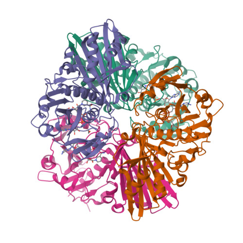
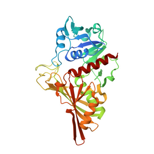
    Glyceraldehyde-3-phosphate dehydrogenase (GAPDH) is a conserved cytosolic enzyme, which plays a key role in glycolysis. GAPDH catalyzes the oxidative phosphorylation of D-glyceraldehyde 3-phosphate using NAD or NADP as a cofactor. In addition, GAPDH localized on the surface of some bacteria is thought to be involved in macromolecular interactions and bacterial pathogenesis. GAPDH on the surface of group B streptococcus (GBS) enhances bacterial virulence and is a potential vaccine candidate. Here, the crystal structure of GBS GAPDH from Streptococcus agalactiae in complex with NAD is reported at 2.46 Å resolution. Although the overall structure of GBS GAPDH is very similar to those of other GAPDHs, the crystal structure reveals a significant difference in the area spanning residues 294-307, which appears to be more acidic. The amino-acid sequence of this region of GBS GAPDH is also distinct compared with other GAPDHs. This region therefore may be of interest as an immunogen for vaccine development.


  • Organizational Affiliation

    Science and Technology Honors Program, University of Alabama at Birmingham, Birmingham, AL 35294, USA.


Macromolecules
Find similar proteins by:  (by identity cutoff)  |  3D Structure
Entity ID: 1
MoleculeChains Sequence LengthOrganismDetailsImage
Glyceraldehyde 3-phosphate dehydrogenase
A, B, C, D
356Streptococcus agalactiae NEM316Mutation(s): 0 
Gene Names: gapCGBS 1811
EC: 1.2.1.12 (PDB Primary Data), 1.2.1 (UniProt)
UniProt
Find proteins for Q8E3E8 (Streptococcus agalactiae serotype III (strain NEM316))
Explore Q8E3E8 
Go to UniProtKB:  Q8E3E8
Entity Groups  
Sequence Clusters30% Identity50% Identity70% Identity90% Identity95% Identity100% Identity
UniProt GroupQ8E3E8
Sequence Annotations
Expand
  • Reference Sequence
Experimental Data & Validation

Experimental Data

  • Method: X-RAY DIFFRACTION
  • Resolution: 2.46 Å
  • R-Value Free:  0.238 (Depositor), 0.240 (DCC) 
  • R-Value Work:  0.204 (Depositor), 0.210 (DCC) 
  • R-Value Observed: 0.206 (Depositor) 
Space Group: P 1 21 1
Unit Cell:
Length ( Å )Angle ( ˚ )
a = 67.771α = 90
b = 107.855β = 106.57
c = 90.931γ = 90
Software Package:
Software NamePurpose
REFMACrefinement
PDB_EXTRACTdata extraction
RAPDdata collection
XDSdata reduction
SCALAdata scaling
PHASERphasing

Structure Validation

View Full Validation Report


Ligand Structure Quality Assessment 

Created with Raphaël 2.3.0Worse 01 BetterLigand structure goodness of fit to experimental dataBest fitted NADClick on this verticalbar to view details

Entry History 

Deposition Data

Revision History  (Full details and data files)

  • Version 1.0: 2014-10-15
    Type: Initial release
  • Version 1.1: 2014-12-24
    Changes: Database references
  • Version 1.2: 2015-01-28
    Changes: Advisory
  • Version 1.3: 2024-02-28
    Changes: Data collection, Database references, Derived calculations
  • Version 1.4: 2024-04-03
    Changes: Refinement description