5DO6 | pdb_00005do6

Crystal structure of Dendroaspis polylepis venom mambalgin-1 T23A mutant


Experimental Data Snapshot

  • Method: X-RAY DIFFRACTION
  • Resolution: 1.70 Å
  • R-Value Free: 
    0.205 (Depositor), 0.200 (DCC) 
  • R-Value Work: 
    0.172 (Depositor), 0.170 (DCC) 
  • R-Value Observed: 
    0.173 (Depositor) 

wwPDB Validation   3D Report Full Report


This is version 1.3 of the entry. See complete history


Literature

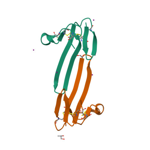
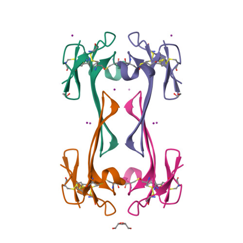
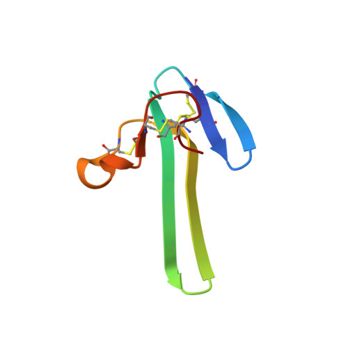
Mambalgin-1 Pain-relieving Peptide, Stepwise Solid-phase Synthesis, Crystal Structure, and Functional Domain for Acid-sensing Ion Channel 1a Inhibition.

Mourier, G.Salinas, M.Kessler, P.Stura, E.A.Leblanc, M.Tepshi, L.Besson, T.Diochot, S.Baron, A.Douguet, D.Lingueglia, E.Servent, D.

(2016) J Biological Chem 291: 2616-2629

  • DOI: https://doi.org/10.1074/jbc.M115.702373
  • Primary Citation of Related Structures:  
    5DO6, 5DU1, 5DZ5

  • PubMed Abstract: 

    Mambalgins are peptides isolated from mamba venom that specifically inhibit a set of acid-sensing ion channels (ASICs) to relieve pain. We show here the first full stepwise solid phase peptide synthesis of mambalgin-1 and confirm the biological activity of the synthetic toxin both in vitro and in vivo. We also report the determination of its three-dimensional crystal structure showing differences with previously described NMR structures. Finally, the functional domain by which the toxin inhibits ASIC1a channels was identified in its loop II and more precisely in the face containing Phe-27, Leu-32, and Leu-34 residues. Moreover, proximity between Leu-32 in mambalgin-1 and Phe-350 in rASIC1a was proposed from double mutant cycle analysis. These data provide information on the structure and on the pharmacophore for ASIC channel inhibition by mambalgins that could have therapeutic value against pain and probably other neurological disorders.


  • Organizational Affiliation

    From the Commissariat à l'Energie Atomique, iBiTecS, Service d'Ingénierie Moléculaire des Protéines, 91191 Gif-sur-Yvette.


Macromolecules
Find similar proteins by:  (by identity cutoff)  |  3D Structure
Entity ID: 1
MoleculeChains Sequence LengthOrganismDetailsImage
Mambalgin-1
A, B
57Dendraspis polylepis polylepisMutation(s): 1 
UniProt
Find proteins for P0DKR6 (Dendroaspis polylepis polylepis)
Explore P0DKR6 
Go to UniProtKB:  P0DKR6
Entity Groups  
Sequence Clusters30% Identity50% Identity70% Identity90% Identity95% Identity100% Identity
UniProt GroupP0DKR6
Sequence Annotations
Expand
  • Reference Sequence
Small Molecules
Ligands 3 Unique
IDChains Name / Formula / InChI Key2D Diagram3D Interactions
IOD
Query on IOD

Download Ideal Coordinates CCD File 
C [auth A],
D [auth A],
E [auth A],
F [auth A],
H [auth B]
IODIDE ION
I
XMBWDFGMSWQBCA-UHFFFAOYSA-M
PGO
Query on PGO

Download Ideal Coordinates CCD File 
I [auth B]S-1,2-PROPANEDIOL
C3 H8 O2
DNIAPMSPPWPWGF-VKHMYHEASA-N
EDO
Query on EDO

Download Ideal Coordinates CCD File 
G [auth A]1,2-ETHANEDIOL
C2 H6 O2
LYCAIKOWRPUZTN-UHFFFAOYSA-N
Experimental Data & Validation

Experimental Data

  • Method: X-RAY DIFFRACTION
  • Resolution: 1.70 Å
  • R-Value Free:  0.205 (Depositor), 0.200 (DCC) 
  • R-Value Work:  0.172 (Depositor), 0.170 (DCC) 
  • R-Value Observed: 0.173 (Depositor) 
Space Group: C 2 2 21
Unit Cell:
Length ( Å )Angle ( ˚ )
a = 55.9α = 90
b = 100.99β = 90
c = 53.48γ = 90
Software Package:
Software NamePurpose
PHENIXrefinement
XDSdata reduction
XSCALEdata scaling
PHENIXphasing
Cootmodel building

Structure Validation

View Full Validation Report


Entry History 

Deposition Data

Revision History  (Full details and data files)

  • Version 1.0: 2015-12-30
    Type: Initial release
  • Version 1.1: 2016-02-17
    Changes: Database references
  • Version 1.2: 2017-01-25
    Changes: Database references
  • Version 1.3: 2024-10-23
    Changes: Data collection, Database references, Structure summary