7E4S | pdb_00007e4s

Crystal structure of Lactobacillus rhamnosus 4-deoxy-L-threo-5-hexosulose-uronate ketol-isomerase KduI complexed with HEPES


Experimental Data Snapshot

  • Method: X-RAY DIFFRACTION
  • Resolution: 2.79 Å
  • R-Value Free: 
    0.238 (Depositor), 0.240 (DCC) 
  • R-Value Work: 
    0.165 (Depositor), 0.170 (DCC) 
  • R-Value Observed: 
    0.168 (Depositor) 

Starting Model: experimental
View more details

wwPDB Validation   3D Report Full Report


Ligand Structure Quality Assessment 

Created with Raphaël 2.3.0Worse 01 BetterLigand structure goodness of fit to experimental dataBest fitted EPEClick on this verticalbar to view details

This is version 1.2 of the entry. See complete history


Literature

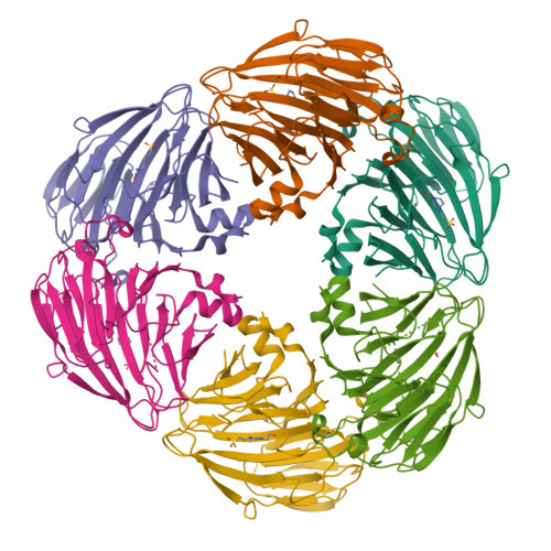
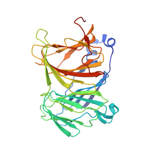
Crystal structures of Lacticaseibacillus 4-deoxy-L-threo-5-hexosulose-uronate ketol-isomerase KduI in complex with substrate analogs

Iwase, H.Yamamoto, Y.Yamada, A.Kawai, K.Oiki, S.Watanabe, D.Mikami, B.Takase, R.Hashimoto, W.

(2023) J Appl Glycosci (1999) 


Macromolecules
Find similar proteins by:  (by identity cutoff)  |  3D Structure
Entity ID: 1
MoleculeChains Sequence LengthOrganismDetailsImage
5-dehydro-4-deoxy-D-glucuronate isomerase
A, B, C, D, E
289Lacticaseibacillus rhamnosusMutation(s): 0 
Gene Names: kduI1kduIGKD15_03440LRHP344_00167
EC: 5.3.1.17
Entity Groups  
Sequence Clusters30% Identity50% Identity70% Identity90% Identity95% Identity100% Identity
Sequence Annotations
Expand
  • Reference Sequence
Small Molecules
Ligands 2 Unique
IDChains Name / Formula / InChI Key2D Diagram3D Interactions
EPE (Subject of Investigation/LOI)
Query on EPE

Download Ideal Coordinates CCD File 
G [auth A]
I [auth B]
K [auth C]
M [auth D]
O [auth E]
4-(2-HYDROXYETHYL)-1-PIPERAZINE ETHANESULFONIC ACID
C8 H18 N2 O4 S
JKMHFZQWWAIEOD-UHFFFAOYSA-N
ZN
Query on ZN

Download Ideal Coordinates CCD File 
H [auth A]
J [auth B]
L [auth C]
N [auth D]
P [auth E]
ZINC ION
Zn
PTFCDOFLOPIGGS-UHFFFAOYSA-N
Experimental Data & Validation

Experimental Data

  • Method: X-RAY DIFFRACTION
  • Resolution: 2.79 Å
  • R-Value Free:  0.238 (Depositor), 0.240 (DCC) 
  • R-Value Work:  0.165 (Depositor), 0.170 (DCC) 
  • R-Value Observed: 0.168 (Depositor) 
Space Group: C 1 2 1
Unit Cell:
Length ( Å )Angle ( ˚ )
a = 94.29α = 90
b = 189.25β = 106.24
c = 117.8γ = 90
Software Package:
Software NamePurpose
PHENIXrefinement
XDSdata scaling
XDSdata reduction
MOLREPphasing

Structure Validation

View Full Validation Report


Ligand Structure Quality Assessment 

Created with Raphaël 2.3.0Worse 01 BetterLigand structure goodness of fit to experimental dataBest fitted EPEClick on this verticalbar to view details

Entry History & Funding Information

Deposition Data


Funding OrganizationLocationGrant Number
Japan Society for the Promotion of Science (JSPS)Japan18H02166

Revision History  (Full details and data files)

  • Version 1.0: 2022-02-23
    Type: Initial release
  • Version 1.1: 2023-11-15
    Changes: Data collection, Database references
  • Version 1.2: 2023-11-29
    Changes: Refinement description