8ZD5 | pdb_00008zd5

Crystal structure of E40K variant of Cu/Zn-superoxide dismutase from dog (Canis familiaris) in the holo form complexed with 22E1 Fv-clasp


Experimental Data Snapshot

  • Method: X-RAY DIFFRACTION
  • Resolution: 2.89 Å
  • R-Value Free: 
    0.249 (Depositor), 0.246 (DCC) 
  • R-Value Work: 
    0.201 (Depositor), 0.204 (DCC) 
  • R-Value Observed: 
    0.203 (Depositor) 

Starting Models: in silico, experimental
View more details

wwPDB Validation   3D Report Full Report


This is version 1.1 of the entry. See complete history


Literature

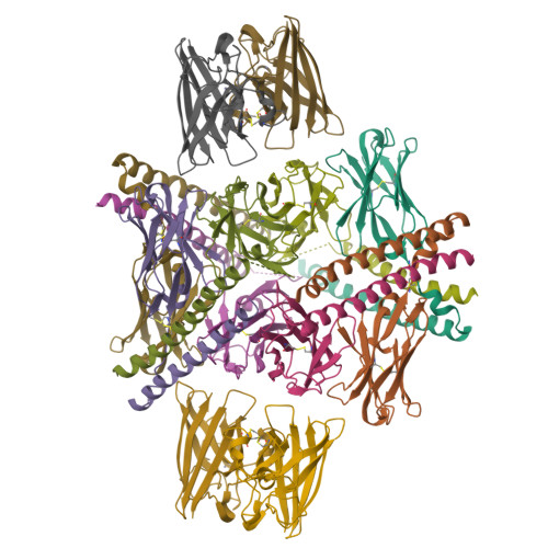
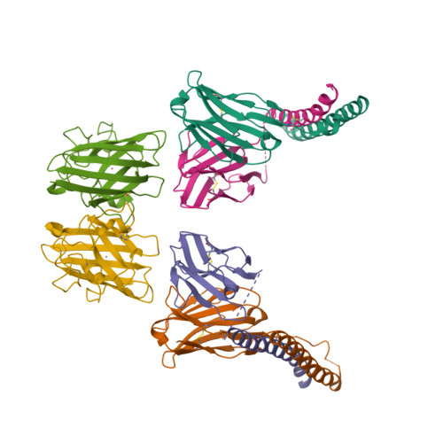
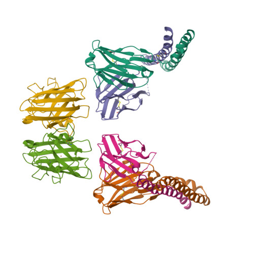
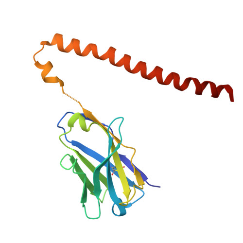
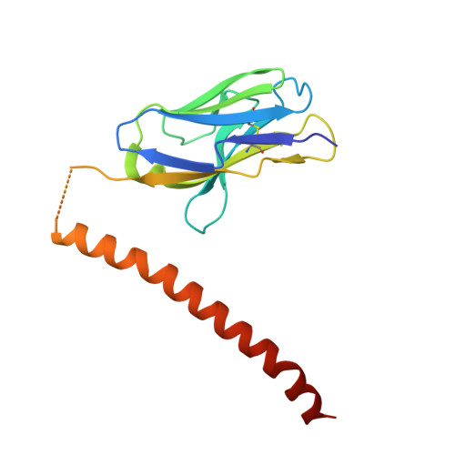
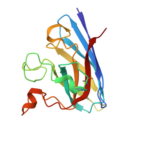
Disulfide-mediated oligomerization of mutant Cu/Zn-superoxide dismutase associated with canine degenerative myelopathy.

Shino, Y.Muraki, N.Kobatake, Y.Kamishina, H.Kato, R.Furukawa, Y.

(2024) Protein Sci 33: e5210-e5210

  • DOI: https://doi.org/10.1002/pro.5210
  • Primary Citation of Related Structures:  
    8ZD5, 8ZD6

  • PubMed Abstract: 

    A homozygous E40K mutation in the gene coding canine Cu/Zn-superoxide dismutase (cSOD1) causes degenerative myelopathy (DM) in dogs. A pathological hallmark of DM with the cSOD1 mutation is the aggregation of mutant cSOD1 proteins in neurons. The amino acid substitution E40K disrupts a salt bridge between Glu40 and Lys91 and is considered to destabilize the native state of cSOD1; however, the mechanism by which mutant cSOD1 aggregates remains unclear. Here, we show that mutant cSOD1 losing a copper and zinc ion forms oligomers crosslinked via disulfide bonds. The E40K substitution was found to result in the increased solvent exposure of the Cys7 side chain, which then attacked the disulfide bond (Cys57-Cys146) in cSOD1 to form disulfide-linked oligomers. We also successfully prevented the Cys7 exposure and thus the oligomerization of mutant cSOD1 by a fragment antibody that specifically recognizes the region around the mutation site. The fragment antibody covered the β-plug region, reinforcing the interactions compromised by the E40K substitution and thus contributing to the maintenance of the structural integrity of the β-barrel core of cSOD1. Taken together, we propose that the Cys7 exposure in cSOD1 upon the salt bridge disruption plays a central role in the aggregation mechanism of DM-associated mutant cSOD1.


  • Organizational Affiliation

    Department of Chemistry, Keio University, Yokohama, Japan.


Macromolecules
Find similar proteins by:  (by identity cutoff)  |  3D Structure
Entity ID: 1
MoleculeChains Sequence LengthOrganismDetailsImage
22E1VH-SARAHA [auth H],
C [auth E],
H [auth B],
K
169Mus musculusMutation(s): 0 
UniProt
Find proteins for A0A075B5S3 (Mus musculus)
Explore A0A075B5S3 
Go to UniProtKB:  A0A075B5S3
Entity Groups  
Sequence Clusters30% Identity50% Identity70% Identity90% Identity95% Identity100% Identity
UniProt GroupA0A075B5S3
Sequence Annotations
Expand
  • Reference Sequence
Find similar proteins by:  (by identity cutoff)  |  3D Structure
Entity ID: 2
MoleculeChains Sequence LengthOrganismDetailsImage
22E1VL-SARAHB [auth F],
I [auth C],
J [auth L],
L [auth I]
159Mus musculusMutation(s): 0 
UniProt
Find proteins for A0A075B5K0 (Mus musculus)
Explore A0A075B5K0 
Go to UniProtKB:  A0A075B5K0
Entity Groups  
Sequence Clusters30% Identity50% Identity70% Identity90% Identity95% Identity100% Identity
UniProt GroupA0A075B5K0
Sequence Annotations
Expand
  • Reference Sequence
Find similar proteins by:  (by identity cutoff)  |  3D Structure
Entity ID: 3
MoleculeChains Sequence LengthOrganismDetailsImage
Superoxide dismutase [Cu-Zn]D [auth A],
E [auth D],
F [auth G],
G [auth J]
153Canis lupus familiarisMutation(s): 1 
Gene Names: SOD1
EC: 1.15.1.1
UniProt
Find proteins for Q8WNN6 (Canis lupus familiaris)
Explore Q8WNN6 
Go to UniProtKB:  Q8WNN6
Entity Groups  
Sequence Clusters30% Identity50% Identity70% Identity90% Identity95% Identity100% Identity
UniProt GroupQ8WNN6
Sequence Annotations
Expand
  • Reference Sequence
Small Molecules
Ligands 2 Unique
IDChains Name / Formula / InChI Key2D Diagram3D Interactions
ZN
Query on ZN

Download Ideal Coordinates CCD File 
M [auth A],
O [auth D],
Q [auth G],
S [auth J]
ZINC ION
Zn
PTFCDOFLOPIGGS-UHFFFAOYSA-N
CU
Query on CU

Download Ideal Coordinates CCD File 
N [auth A],
P [auth D],
R [auth G],
T [auth J]
COPPER (II) ION
Cu
JPVYNHNXODAKFH-UHFFFAOYSA-N
Experimental Data & Validation

Experimental Data

  • Method: X-RAY DIFFRACTION
  • Resolution: 2.89 Å
  • R-Value Free:  0.249 (Depositor), 0.246 (DCC) 
  • R-Value Work:  0.201 (Depositor), 0.204 (DCC) 
  • R-Value Observed: 0.203 (Depositor) 
Space Group: I 1 2 1
Unit Cell:
Length ( Å )Angle ( ˚ )
a = 167.915α = 90
b = 82.213β = 108.46
c = 187.612γ = 90
Software Package:
Software NamePurpose
REFMACrefinement
Aimlessdata scaling
MOLREPphasing
Cootmodel building
XDSdata reduction

Structure Validation

View Full Validation Report


Entry History & Funding Information

Deposition Data


Funding OrganizationLocationGrant Number
Not funded--

Revision History  (Full details and data files)

  • Version 1.0: 2024-11-27
    Type: Initial release
  • Version 1.1: 2024-12-04
    Changes: Database references