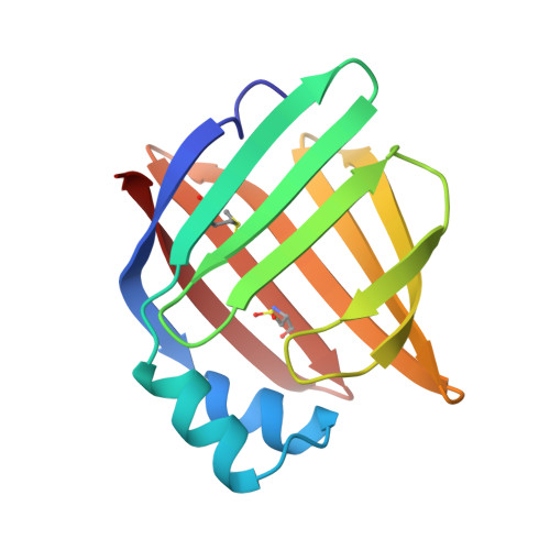
1ADL

ADIPOCYTE LIPID BINDING PROTEIN COMPLEXED WITH ARACHIDONIC ACID: X-RAY CRYSTALLOGRAPHIC AND TITRATION CALORIMETRY STUDIES


Experimental Data Snapshot

  • Method: X-RAY DIFFRACTION
  • Resolution: 1.60 Å
  • R-Value Free: 0.220 
  • R-Value Work: 0.188 
  • R-Value Observed: 0.188 

wwPDB Validation   3D Report Full Report


This is version 2.1 of the entry. See complete history


Literature

Adipocyte lipid-binding protein complexed with arachidonic acid. Titration calorimetry and X-ray crystallographic studies.

LaLonde, J.M.Levenson, M.A.Roe, J.J.Bernlohr, D.A.Banaszak, L.J.

(1994) J Biol Chem 269: 25339-25347

  • DOI: https://doi.org/10.2210/pdb1adl/pdb
  • Primary Citation of Related Structures:  
    1ADL

  • PubMed Abstract: 

    The association of the adipocyte lipid-binding protein (ALBP) with arachidonic acid (all cis, 20:4 delta 5,8,11,14) and oleic acid (cis, 18:1 delta 9) has been examined by titration calorimentry. In addition, the crystal structure of ALBP with bound arachidonic acid has also been obtained. Crystallographic analysis of the arachidonic acid.ALBP complex along with the previously reported oleic acid-ALBP structure (Xu, Z., Bernlohr, D. A., and Banaszak, L. J. (1993) J. Biol. Chem. 268, 7874-7884) provides a framework for the molecular examination of protein-lipid association. Isothermal titration calorimetry revealed high affinity association of both unsaturated fatty acids with the protein. The calorimetric data yielded the following thermodynamic parameters for arachidonic acid: Kd = 4.4 microM, n = 0.8, delta G = -7370 cal/mol, delta H = -6770 cal/mol, and T delta S = +600 cal/mol. For oleic acid, the thermodynamic parameters were Kd = 2.4 microM, n = 0.9, delta G = -7770 cal/mol, delta H = -6050 cal/mol, and T delta S = +1720 cal/mol. The identification of thermodynamically dominating enthalpic factors for both fatty acids are consistent with the crystallographic studies demonstrating the interaction of the fatty acid carboxylate with a combination of Arg106, Arg126, and Tyr128. The crystallographic refinement of the protein-arachidonate complex was carried out to 1.6 A with the resultant R factor of 0.19. Within the cavity of the crystalline binding protein, the arachidonate was found in a hairpin conformation. The conformation of the bound ligand is consistent with acceptable torsional angles and the four cis double bonds in arachidonate. These results demonstrate that arachidonate is a ligand for ALBP. They provide thermodynamic and structural data concerning the physical basis for protein-lipid interaction and suggest that intracellular lipid-binding proteins may mediate the biological effects of polyunsaturated fatty acids in vivo.


  • Organizational Affiliation

    Department of Biochemistry, School of Medicine, University of Minnesota, Minneapolis 55455.


Macromolecules
Find similar proteins by:  (by identity cutoff)  |  3D Structure
Entity ID: 1
MoleculeChains Sequence LengthOrganismDetailsImage
ADIPOCYTE LIPID-BINDING PROTEIN131Mus musculusMutation(s): 0 
UniProt
Find proteins for P04117 (Mus musculus)
Explore P04117 
Go to UniProtKB:  P04117
Entity Groups  
Sequence Clusters30% Identity50% Identity70% Identity90% Identity95% Identity100% Identity
UniProt GroupP04117
Sequence Annotations
Expand
  • Reference Sequence
Small Molecules
Ligands 2 Unique
IDChains Name / Formula / InChI Key2D Diagram3D Interactions
ACD
Query on ACD

Download Ideal Coordinates CCD File 
B [auth A]ARACHIDONIC ACID
C20 H32 O2
YZXBAPSDXZZRGB-DOFZRALJSA-N
PPI
Query on PPI

Download Ideal Coordinates CCD File 
C [auth A]PROPANOIC ACID
C3 H6 O2
XBDQKXXYIPTUBI-UHFFFAOYSA-N
Modified Residues  2 Unique
IDChains TypeFormula2D DiagramParent
CSD
Query on CSD
A
L-PEPTIDE LINKINGC3 H7 N O4 SCYS
MHO
Query on MHO
A
L-PEPTIDE LINKINGC5 H11 N O3 SMET
Binding Affinity Annotations 
IDSourceBinding Affinity
ACD BindingDB:  1ADL Kd: 180 (nM) from 1 assay(s)
IC50: min: 2750, max: 5130 (nM) from 2 assay(s)
PDBBind:  1ADL Kd: 4400 (nM) from 1 assay(s)
Experimental Data & Validation

Experimental Data

  • Method: X-RAY DIFFRACTION
  • Resolution: 1.60 Å
  • R-Value Free: 0.220 
  • R-Value Work: 0.188 
  • R-Value Observed: 0.188 
  • Space Group: C 1 2 1
Unit Cell:
Length ( Å )Angle ( ˚ )
a = 120.54α = 90
b = 37.7β = 92.34
c = 28.57γ = 90
Software Package:
Software NamePurpose
X-PLORmodel building
X-PLORrefinement
X-PLORphasing

Structure Validation

View Full Validation Report


Entry History 

Deposition Data

Revision History  (Full details and data files)

  • Version 1.0: 1994-12-20
    Type: Initial release
  • Version 1.1: 2008-03-24
    Changes: Version format compliance
  • Version 1.2: 2011-07-13
    Changes: Version format compliance
  • Version 1.3: 2017-11-29
    Changes: Advisory, Derived calculations, Other, Structure summary
  • Version 2.0: 2024-02-07
    Changes: Advisory, Atomic model, Data collection, Database references, Derived calculations, Non-polymer description, Polymer sequence, Source and taxonomy, Structure summary
  • Version 2.1: 2024-10-09
    Changes: Structure summary