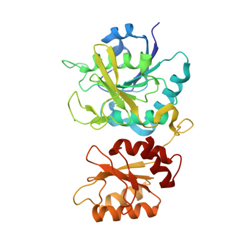
Stuctural Basis for the Activity and Substrate Specificity of Erwinia Chrysanthemi L-Asparaginase
Kolyani, K.A., Wlodawer, A., Lubkowski, J.(2001) Biochemistry 40: 5655
- PubMed: 11341830
- DOI: https://doi.org/10.1021/bi0029595
- Primary Citation of Related Structures:
1HFW, 1HG0, 1HG1 - PubMed Abstract:
Bacterial L-asparaginases, enzymes that catalyze the hydrolysis of L-asparagine to aspartic acid, have been used for over 30 years as therapeutic agents in the treatment of acute childhood lymphoblastic leukemia. Other substrates of asparaginases include L-glutamine, D-asparagine, and succinic acid monoamide. In this report, we present high-resolution crystal structures of the complexes of Erwinia chrysanthemi L-asparaginase (ErA) with the products of such reactions that also can serve as substrates, namely L-glutamic acid (L-Glu), D-aspartic acid (D-Asp), and succinic acid (Suc). Comparison of the four independent active sites within each complex indicates unique and specific binding of the ligand molecules; the mode of binding is also similar between complexes. The lack of the alpha-NH3(+) group in Suc, compared to L-Asp, does not affect the binding mode. The side chain of L-Glu, larger than that of L-Asp, causes several structural distortions in the ErA active side. The active site flexible loop (residues 15-33) does not exhibit stable conformation, resulting in suboptimal orientation of the nucleophile, Thr15. Additionally, the delta-COO(-) plane of L-Glu is approximately perpendicular to the plane of gamma-COO(-) in L-Asp bound to the asparaginase active site. Binding of D-Asp to the ErA active site is very distinctive compared to the other ligands, suggesting that the low activity of ErA against D-Asp could be mainly attributed to the low k(cat) value. A comparison of the amino acid sequence and the crystal structure of ErA with those of other bacterial L-asparaginases shows that the presence of two active-site residues, Glu63(ErA) and Ser254(ErA), may correlate with significant glutaminase activity, while their substitution by Gln and Asn, respectively, may lead to minimal L-glutaminase activity.
Organizational Affiliation:
Macromolecular Crystallography Laboratory, National Cancer Institute at Frederick, Frederick, Maryland 21702, USA.


