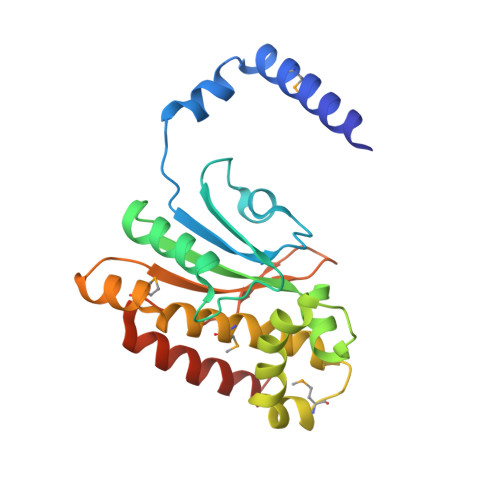
1I6O

CRYSTAL STRUCTURE OF E. COLI BETA CARBONIC ANHYDRASE (ECCA)


Experimental Data Snapshot

  • Method: X-RAY DIFFRACTION
  • Resolution: 2.20 Å
  • R-Value Free: 0.243 
  • R-Value Work: 0.212 
  • R-Value Observed: 0.212 

wwPDB Validation   3D Report Full Report


This is version 1.3 of the entry. See complete history


Literature

Crystal structure of E. coli beta-carbonic anhydrase, an enzyme with an unusual pH-dependent activity.

Cronk, J.D.Endrizzi, J.A.Cronk, M.R.O'neill, J.W.Zhang, K.Y.

(2001) Protein Sci 10: 911-922

  • DOI: https://doi.org/10.1110/ps.46301
  • Primary Citation of Related Structures:  
    1I6O, 1I6P

  • PubMed Abstract: 

    Carbonic anhydrases fall into three distinct evolutionary and structural classes: alpha, beta, and gamma. The beta-class carbonic anhydrases (beta-CAs) are widely distributed among higher plants, simple eukaryotes, eubacteria, and archaea. We have determined the crystal structure of ECCA, a beta-CA from Escherichia coli, to a resolution of 2.0 A. In agreement with the structure of the beta-CA from the chloroplast of the red alga Porphyridium purpureum, the active-site zinc in ECCA is tetrahedrally coordinated by the side chains of four conserved residues. These results confirm the observation of a unique pattern of zinc ligation in at least some beta-CAS: The absence of a water molecule in the inner coordination sphere is inconsistent with known mechanisms of CA activity. ECCA activity is highly pH-dependent in the physiological range, and its expression in yeast complements an oxygen-sensitive phenotype displayed by a beta-CA-deletion strain. The structural and biochemical characterizations of ECCA presented here and the comparisons with other beta-CA structures suggest that ECCA can adopt two distinct conformations displaying widely divergent catalytic rates.


  • Organizational Affiliation

    Division of Basic Sciences, Fred Hutchinson Cancer Research Center, Seattle, Washington 98109-1024, USA.


Macromolecules
Find similar proteins by:  (by identity cutoff)  |  3D Structure
Entity ID: 1
MoleculeChains Sequence LengthOrganismDetailsImage
CARBONIC ANHYDRASE
A, B
220Escherichia coliMutation(s): 5 
Gene Names: YADF
EC: 4.2.1.1
UniProt
Find proteins for P61517 (Escherichia coli (strain K12))
Explore P61517 
Go to UniProtKB:  P61517
Entity Groups  
Sequence Clusters30% Identity50% Identity70% Identity90% Identity95% Identity100% Identity
UniProt GroupP61517
Sequence Annotations
Expand
  • Reference Sequence
Small Molecules
Modified Residues  1 Unique
IDChains TypeFormula2D DiagramParent
MSE
Query on MSE
A, B
L-PEPTIDE LINKINGC5 H11 N O2 SeMET
Experimental Data & Validation

Experimental Data

  • Method: X-RAY DIFFRACTION
  • Resolution: 2.20 Å
  • R-Value Free: 0.243 
  • R-Value Work: 0.212 
  • R-Value Observed: 0.212 
  • Space Group: P 43 2 2
Unit Cell:
Length ( Å )Angle ( ˚ )
a = 81.244α = 90
b = 81.244β = 90
c = 162.137γ = 90
Software Package:
Software NamePurpose
CNSrefinement
MOSFLMdata reduction
CCP4data scaling
CNSphasing

Structure Validation

View Full Validation Report


Entry History 

Deposition Data

Revision History  (Full details and data files)

  • Version 1.0: 2001-05-09
    Type: Initial release
  • Version 1.1: 2008-04-27
    Changes: Version format compliance
  • Version 1.2: 2011-07-13
    Changes: Derived calculations, Version format compliance
  • Version 1.3: 2024-10-16
    Changes: Data collection, Database references, Derived calculations, Structure summary