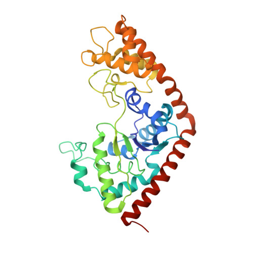
Interconversion of ATP binding and conformational free energies by tryptophanyl-tRNA synthetase: structures of ATP bound to open and closed, pre-transition-state conformations.
Retailleau, P., Huang, X., Yin, Y., Hu, M., Weinreb, V., Vachette, P., Vonrhein, C., Bricogne, G., Roversi, P., Ilyin, V., Carter, C.W.(2003) J Mol Biol 325: 39-63
- PubMed: 12473451
- DOI: https://doi.org/10.1016/s0022-2836(02)01156-7
- Primary Citation of Related Structures:
1M83, 1MAU, 1MAW, 1MB2 - PubMed Abstract:
Binding ATP to tryptophanyl-tRNA synthetase (TrpRS) in a catalytically competent configuration for amino acid activation destabilizes the enzyme structure prior to forming the transition state. This conclusion follows from monitoring the titration of TrpRS with ATP by small angle solution X-ray scattering, enzyme activity, and crystal structures. ATP induces a significantly smaller radius of gyration at pH=7 with a transition midpoint at approximately 8mM. A non-reciprocal dependence of Trp and ATP dissociation constants on concentrations of the second substrate show that Trp binding enhances affinity for ATP, while the affinity for Trp falls with the square of the [ATP] over the same concentration range ( approximately 5mM) that induces the more compact conformation. Two distinct TrpRS:ATP structures have been solved, a high-affinity complex grown with 1mM ATP and a low-affinity complex grown at 10mM ATP. The former is isomorphous with unliganded TrpRS and the Trp complex from monoclinic crystals. Reacting groups of the two individually-bound substrates are separated by 6.7A. Although it lacks tryptophan, the low-affinity complex has a closed conformation similar to that observed in the presence of both ATP and Trp analogs such as indolmycin, and resembles a complex previously postulated to form in the closely-related TyrRS upon induced-fit active-site assembly, just prior to catalysis. Titration of TrpRS with ATP therefore successively produces structurally distinct high- and low-affinity ATP-bound states. The higher quality X-ray data for the closed ATP complex (2.2A) provide new structural details likely related to catalysis, including an extension of the KMSKS loop that engages the second lysine and serine residues, K195 and S196, with the alpha and gamma-phosphates; interactions of the K111 side-chain with the gamma-phosphate; and a water molecule bridging the consensus sequence residue T15 to the beta-phosphate. Induced-fit therefore strengthens active-site interactions with ATP, substantially intensifying the interaction of the KMSKS loop with the leaving PP(i) group. Formation of this conformation in the absence of a Trp analog implies that ATP is a key allosteric effector for TrpRS. The paradoxical requirement for high [ATP] implies that Gibbs binding free energy is stored in an unfavorable protein conformation and can then be recovered for useful purposes, including catalysis in the case of TrpRS.
Organizational Affiliation:
Department of Biochemistry and Biophysics, University of North Carolina, Mary Ellen Jones Bldg. CB# 7260, Chapel Hill, NC 27599-7260, USA.


