1MX0 | pdb_00001mx0

Structure of topoisomerase subunit


Experimental Data Snapshot

  • Method: X-RAY DIFFRACTION
  • Resolution: 2.30 Å
  • R-Value Free: 
    0.263 (Depositor), 0.258 (DCC) 
  • R-Value Work: 
    0.214 (Depositor), 0.216 (DCC) 
  • R-Value Observed: 
    0.219 (Depositor) 

wwPDB Validation   3D Report Full Report


Ligand Structure Quality Assessment 

Created with Raphaël 2.3.0Worse 01 BetterLigand structure goodness of fit to experimental dataBest fitted ANPClick on this verticalbar to view details

This is version 1.3 of the entry. See complete history


Literature

Structure of the topoisomerase VI-B subunit: implications for type II topoisomerase mechanism and evolution

Corbett, K.D.Berger, J.M.

(2003) EMBO J 22: 151-163

  • DOI: https://doi.org/10.1093/emboj/cdg008
  • Primary Citation of Related Structures:  
    1MU5, 1MX0

  • PubMed Abstract: 

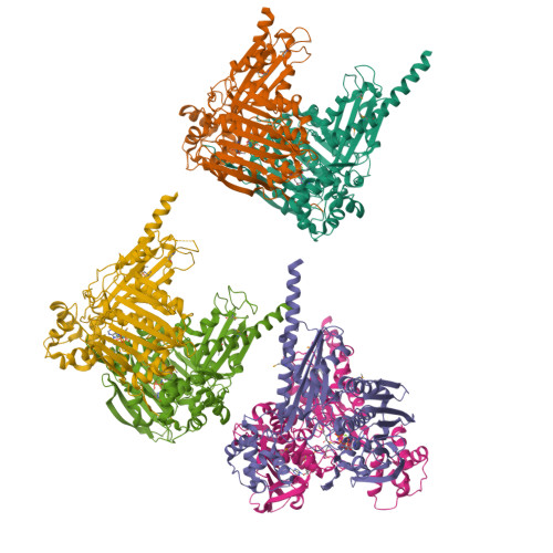
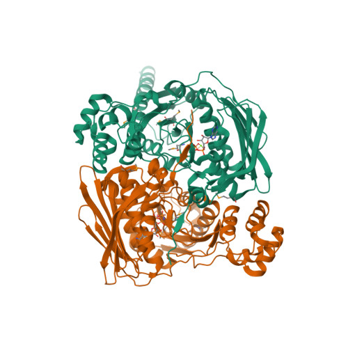
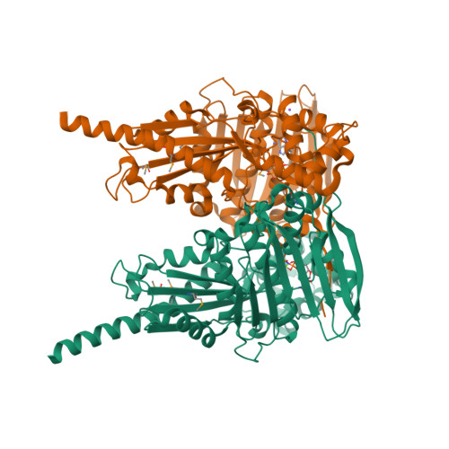
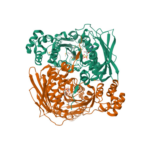
    Type IIA and type IIB topoisomerases each possess the ability to pass one DNA duplex through another in an ATP-dependent manner. The role of ATP in the strand passage reaction is poorly understood, particularly for the type IIB (topoisomerase VI) family. We have solved the structure of the ATP-binding subunit of topoisomerase VI (topoVI-B) in two states: an unliganded monomer and a nucleotide-bound dimer. We find that topoVI-B is highly structurally homologous to the entire 40-43 kDa ATPase region of type IIA topoisomerases and MutL proteins. Nucleotide binding to topoVI-B leads to dimerization of the protein and causes dramatic conformational changes within each protomer. Our data demonstrate that type IIA and type IIB topoisomerases have descended from a common ancestor and reveal how ATP turnover generates structural signals in the reactions of both type II topoisomerase families. When combined with the structure of the A subunit to create a picture of the intact topoisomerase VI holoenzyme, the ATP-driven motions of topoVI-B reveal a simple mechanism for strand passage by the type IIB topoisomerases.


  • Organizational Affiliation

    Department of Molecular and Cellular Biology, University of California, Berkeley, 327 Hildebrand Hall 3206, Berkeley, CA 94720, USA.


Macromolecules
Find similar proteins by:  (by identity cutoff)  |  3D Structure
Entity ID: 1
MoleculeChains Sequence LengthOrganismDetailsImage
Type II DNA topoisomerase VI subunit B
A, B, C, D, E
472Saccharolobus shibataeMutation(s): 5 
Gene Names: top6b
EC: 5.99.1.3 (PDB Primary Data), 5.6.2.2 (UniProt)
UniProt
Find proteins for O05207 (Saccharolobus shibatae (strain ATCC 51178 / DSM 5389 / JCM 8931 / NBRC 15437 / B12))
Explore O05207 
Go to UniProtKB:  O05207
Entity Groups  
Sequence Clusters30% Identity50% Identity70% Identity90% Identity95% Identity100% Identity
UniProt GroupO05207
Sequence Annotations
Expand
  • Reference Sequence
Small Molecules
Ligands 3 Unique
IDChains Name / Formula / InChI Key2D Diagram3D Interactions
ANP
Query on ANP

Download Ideal Coordinates CCD File 
H [auth A]
J [auth B]
L [auth C]
O [auth D]
Q [auth E]
PHOSPHOAMINOPHOSPHONIC ACID-ADENYLATE ESTER
C10 H17 N6 O12 P3
PVKSNHVPLWYQGJ-KQYNXXCUSA-N
MG
Query on MG

Download Ideal Coordinates CCD File 
G [auth A]
I [auth B]
K [auth C]
M [auth D]
P [auth E]
MAGNESIUM ION
Mg
JLVVSXFLKOJNIY-UHFFFAOYSA-N
NA
Query on NA

Download Ideal Coordinates CCD File 
N [auth D]SODIUM ION
Na
FKNQFGJONOIPTF-UHFFFAOYSA-N
Modified Residues  1 Unique
IDChains TypeFormula2D DiagramParent
MSE
Query on MSE
A, B, C, D, E
L-PEPTIDE LINKINGC5 H11 N O2 SeMET
Experimental Data & Validation

Experimental Data

  • Method: X-RAY DIFFRACTION
  • Resolution: 2.30 Å
  • R-Value Free:  0.263 (Depositor), 0.258 (DCC) 
  • R-Value Work:  0.214 (Depositor), 0.216 (DCC) 
  • R-Value Observed: 0.219 (Depositor) 
Space Group: P 21 21 2
Unit Cell:
Length ( Å )Angle ( ˚ )
a = 146.655α = 90
b = 219.192β = 90
c = 106.921γ = 90
Software Package:
Software NamePurpose
DENZOdata reduction
SCALEPACKdata scaling
SHARPphasing
REFMACrefinement

Structure Validation

View Full Validation Report


Ligand Structure Quality Assessment 

Created with Raphaël 2.3.0Worse 01 BetterLigand structure goodness of fit to experimental dataBest fitted ANPClick on this verticalbar to view details

Entry History 

Deposition Data

Revision History  (Full details and data files)

  • Version 1.0: 2003-01-07
    Type: Initial release
  • Version 1.1: 2008-04-28
    Changes: Version format compliance
  • Version 1.2: 2011-07-13
    Changes: Advisory, Version format compliance
  • Version 1.3: 2024-10-16
    Changes: Data collection, Database references, Derived calculations, Structure summary