1N51

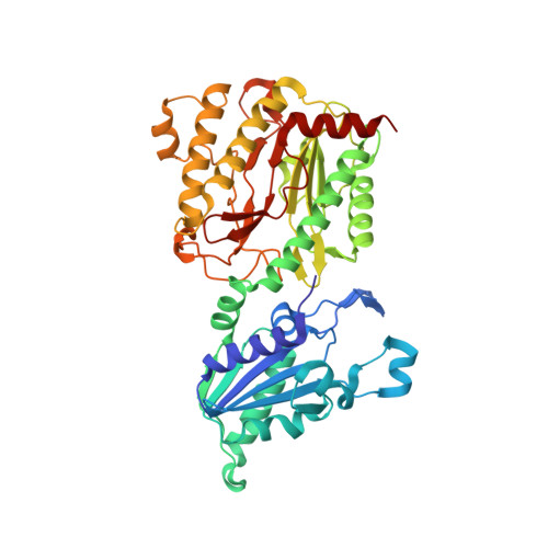
Aminopeptidase P in complex with the inhibitor apstatin


Experimental Data Snapshot

  • Method: X-RAY DIFFRACTION
  • Resolution: 2.30 Å
  • R-Value Free: 0.204 
  • R-Value Work: 0.179 

Starting Model: experimental
View more details

wwPDB Validation   3D Report Full Report


This is version 2.0 of the entry. See complete history


Literature

Structure of Escherichia coli aminopeptidase P in complex with the inhibitor apstatin.

Graham, S.C.Maher, M.J.Simmons, W.H.Freeman, H.C.Guss, J.M.

(2004) Acta Crystallogr D Biol Crystallogr 60: 1770-1779

  • DOI: https://doi.org/10.1107/S0907444904018724
  • Primary Citation of Related Structures:  
    1N51

  • PubMed Abstract: 

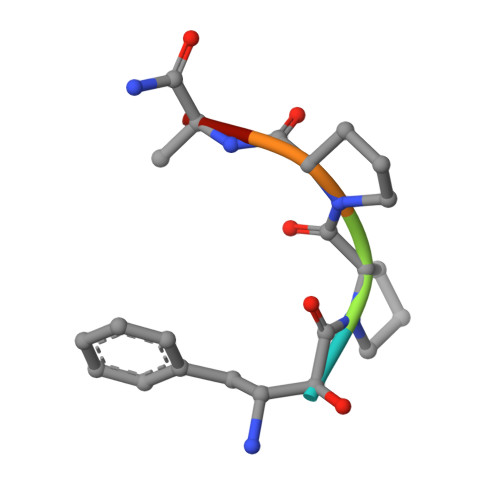
    Aminopeptidase P (APPro) is a metalloprotease whose active site includes a dinuclear manganese(II) cluster. The enzyme cleaves the N-terminal residue from a polypeptide when the second residue is proline. A complex of Escherichia coli APPro (EcAPPro) with an inhibitor, apstatin [N-(2S,3R)-3-amino-2-hydroxy-4-phenyl-butanoyl-L-prolyl-L-prolyl-L-alaninamide], has been crystallized. Apstatin binds to the active site of EcAPPro with its N-terminal amino group coordinated to one of the two Mn(II) atoms at the metal centre. The apstatin hydroxyl group replaces a hydroxide ion which bridges the two metal atoms in the native enzyme. The first proline residue of apstatin lies in a small hydrophobic cleft. The structure of the apstatin-EcAPPro complex has been refined at 2.3 A resolution with residuals R = 0.179 and R(free) = 0.204. The structure of the complex illustrates how apstatin inhibits APPro and suggests how substrates may bind to the enzyme, but the basis of the proline-specificity remains elusive.


  • Organizational Affiliation

    School of Molecular and Microbial Biosciences, University of Sydney, NSW 2006, Australia.


Macromolecules
Find similar proteins by:  (by identity cutoff)  |  3D Structure
Entity ID: 1
MoleculeChains Sequence LengthOrganismDetailsImage
Xaa-Pro aminopeptidase440Escherichia coliMutation(s): 0 
Gene Names: PEPP
EC: 3.4.11.9
UniProt
Find proteins for P15034 (Escherichia coli (strain K12))
Explore P15034 
Go to UniProtKB:  P15034
Entity Groups  
Sequence Clusters30% Identity50% Identity70% Identity90% Identity95% Identity100% Identity
UniProt GroupP15034
Sequence Annotations
Expand
  • Reference Sequence

Find similar proteins by:  Sequence   |   3D Structure  

Entity ID: 2
MoleculeChains Sequence LengthOrganismDetailsImage
apstatin5N/AMutation(s): 0 
Sequence Annotations
Expand
  • Reference Sequence
Biologically Interesting Molecules (External Reference) 1 Unique
Experimental Data & Validation

Experimental Data

  • Method: X-RAY DIFFRACTION
  • Resolution: 2.30 Å
  • R-Value Free: 0.204 
  • R-Value Work: 0.179 
  • Space Group: I 41 2 2
Unit Cell:
Length ( Å )Angle ( ˚ )
a = 139.319α = 90
b = 139.319β = 90
c = 231.005γ = 90
Software Package:
Software NamePurpose
CNSrefinement
DENZOdata reduction
SCALEPACKdata scaling
AMoREphasing

Structure Validation

View Full Validation Report


Entry History 

Deposition Data

Revision History  (Full details and data files)

  • Version 1.0: 2003-12-16
    Type: Initial release
  • Version 1.1: 2008-04-28
    Changes: Version format compliance
  • Version 1.2: 2011-07-13
    Changes: Atomic model, Database references, Derived calculations, Non-polymer description, Structure summary, Version format compliance
  • Version 1.3: 2012-12-12
    Changes: Other
  • Version 1.4: 2023-10-25
    Changes: Data collection, Database references, Derived calculations, Refinement description
  • Version 2.0: 2023-11-15
    Changes: Atomic model, Data collection, Derived calculations