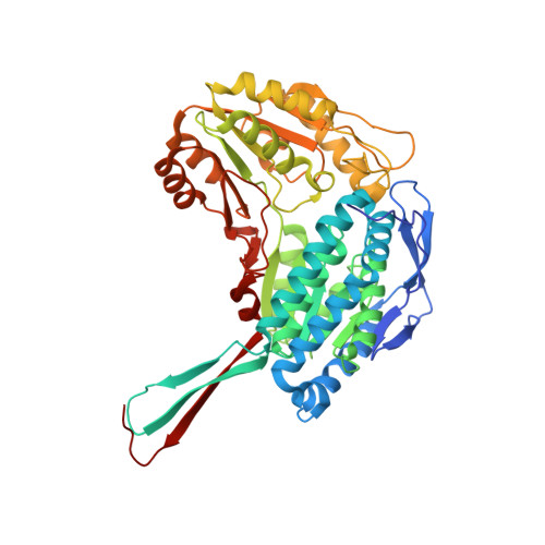
Order and disorder in mitochondrial aldehyde dehydrogenase
Hurley, T.D., Perez-Miller, S., Breen, H.(2001) Chem Biol Interact 130-132: 3-14
- PubMed: 11306026
- DOI: https://doi.org/10.1016/s0009-2797(00)00217-9
- Primary Citation of Related Structures:
1O05 - PubMed Abstract:
One of the most notable and currently unexplained features of the mitochondrial form of aldehyde dehydrogenase is its property of half-of-the-sites reactivity. An appropriate description of this phenomenon can be to consider this as the extreme example of negative cooperativity. This implies, therefore, that a pathway of communication must exist between active sites in order to convey the structural consequences of ligand binding. Data from four different structures of human ALDH2 collected during the past 2 years may shed some light on one possible pathway for the propagation of structural information. We recently published a 2.6 A structure of a binary complex between ALDH2 and NAD(+) in which the predominant conformation of the cofactor differed between different subunits in the structure. We now have three unpublished structures, a wild-type apo-enzyme structure at 2.25 A resolution, a wild-type structure complexed with NADH at 2.45 A resolution, and a site-directed mutant of ALDH2 where Arg475 is mutated to Gln, as an apo-enzyme to 2.75 A resolution. A detailed comparison of their structures reveals that a disorder-to-order transition occurs upon coenzyme binding in the area immediately surrounding the adenosine-binding site (residues 224-233 and 246-262). These residues correspond to the two helices that surround the adenine ring of the cofactor. Since the helix comprised of residues 246-262 contacts its dimer related helix across the subunit interface, this could induce as of yet unidentified subtle changes in structure that impair productive binding of the cofactor in the second subunit. The unique characteristics and three-dimensional structure of the R475Q variant of ALDH2 supports a role in subunit communication for these residues. This mutated enzyme displays positive cooperativity for cofactor binding. The structure of the apo-enzyme shows that the average thermal parameters for the residues involved in adenosine binding are drastically elevated as is a stretch of amino acids surrounding the site of mutation (residues 471-480). We hypothesize that cofactor binding displays a Hill coefficient of approximately 2 because binding of coenzyme to one subunit in a dimer orders the residues responsible for cofactor binding in the second, thus promoting binding. The difference between these alterations being positively versus negatively cooperative is likely related to the magnitude of the structural changes. Further work is in progress to confirm this hypothesis as it may shed light on the dominant effects of the E487K allelic variant, since Glu487 interacts with Arg475.
Organizational Affiliation:
Department of Biochemistry and Molecular Biology, Program in Medical Biophysics, Indiana University School of Medicine, 635 Barnhill Drive, 46202-5122, Indianapolis, IN, USA. thurley@iupui.edu


