1P06

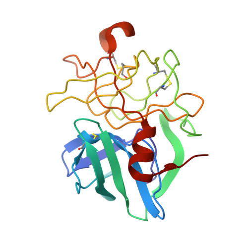
STRUCTURE ANALYSIS OF SPECIFICITY. ALPHA-LYTIC PROTEASE COMPLEXES WITH ANALOGUES OF REACTION INTERMEDIATES


Experimental Data Snapshot

  • Method: X-RAY DIFFRACTION
  • Resolution: 2.34 Å
  • R-Value Observed: 0.140 

wwPDB Validation   3D Report Full Report


This is version 1.5 of the entry. See complete history


Literature

Structural analysis of specificity: alpha-lytic protease complexes with analogues of reaction intermediates.

Bone, R.Frank, D.Kettner, C.A.Agard, D.A.

(1989) Biochemistry 28: 7600-7609

  • DOI: https://doi.org/10.1021/bi00445a015
  • Primary Citation of Related Structures:  
    1P02, 1P03, 1P04, 1P05, 1P06

  • PubMed Abstract: 

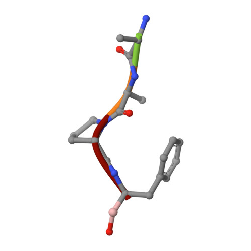
    To better understand the structural basis of enzyme specificity, the structures of complexes formed between alpha-lytic protease, an extracellular serine protease of Lysobacter enzymogenes, and five inhibitory peptide boronic acids (R2-boroX, where R2 is methoxysuccinyl-Ala-Ala-Pro- and boroX is the alpha-aminoboronic acid analogue of Ala, Val, Ile, Norleu, or Phe) have been studied at high resolution by X-ray crystallography. The enzyme has primary specificity for Ala in the P1 position of peptide substrates with catalytic efficiency decreasing with increasing side-chain volume. Enzyme affinity for inhibitors with boroVal, boroIle, and boroPhe residues is much higher than expected on the basis of the catalytic efficiencies of homologous substrates. Covalent tetrahedral adducts are formed between the active-site serine and the boronic acid moieties of R2-boroAla, R2-boroVal R2-boroIle, and R2-boroNorleu. Though R2-boroVal is a slowly bound inhibitor and R2-boroAla is rapidly bound [Kettner, C. A., Bone, R., Agard, D. A., & Bachovchin, W. W. (1988) Biochemistry 27, 7682-7688], there appear to be no structural differences that could account for slow binding. The removal from solution of 20% more hydrophobic surface on binding accounts for the improved affinity of alpha-lytic protease for R2-boroVal relative to R2-boroAla. The high affinity of the enzyme for R2-boroIle derives from the selective binding of the L-allo stereoisomer of the boroIle residue, which can avoid bad steric interactions in the binding pocket.(ABSTRACT TRUNCATED AT 250 WORDS)


  • Organizational Affiliation

    Department of Biochemistry and Biophysics, University of California, San Francisco 94143-0448.


Macromolecules
Find similar proteins by:  (by identity cutoff)  |  3D Structure
Entity ID: 1
MoleculeChains Sequence LengthOrganismDetailsImage
ALPHA-LYTIC PROTEASE198Lysobacter enzymogenesMutation(s): 0 
EC: 3.4.21.12
UniProt
Find proteins for P00778 (Lysobacter enzymogenes)
Explore P00778 
Go to UniProtKB:  P00778
Entity Groups  
Sequence Clusters30% Identity50% Identity70% Identity90% Identity95% Identity100% Identity
UniProt GroupP00778
Sequence Annotations
Expand
  • Reference Sequence

Find similar proteins by:  Sequence   |   3D Structure  

Entity ID: 2
MoleculeChains Sequence LengthOrganismDetailsImage
METHOXYSUCCINYL-ALA-ALA-PRO-PHENYLALANINE BORONIC ACID INHIBITORB [auth P]5N/AMutation(s): 0 
Sequence Annotations
Expand
  • Reference Sequence
Small Molecules
Ligands 1 Unique
IDChains Name / Formula / InChI Key2D Diagram3D Interactions
SO4
Query on SO4

Download Ideal Coordinates CCD File 
C [auth A]SULFATE ION
O4 S
QAOWNCQODCNURD-UHFFFAOYSA-L
Modified Residues  1 Unique
IDChains TypeFormula2D DiagramParent
B2F
Query on B2F
B [auth P]PEPTIDE-LIKEC8 H12 B N O2PHE
Biologically Interesting Molecules (External Reference) 1 Unique
Experimental Data & Validation

Experimental Data

  • Method: X-RAY DIFFRACTION
  • Resolution: 2.34 Å
  • R-Value Observed: 0.140 
  • Space Group: P 32 2 1
Unit Cell:
Length ( Å )Angle ( ˚ )
a = 66.62α = 90
b = 66.62β = 90
c = 80.22γ = 120
Software Package:
Software NamePurpose
PROLSQrefinement

Structure Validation

View Full Validation Report


Entry History 

Deposition Data

Revision History  (Full details and data files)

  • Version 1.0: 1990-04-15
    Type: Initial release
  • Version 1.1: 2008-03-25
    Changes: Version format compliance
  • Version 1.2: 2011-07-13
    Changes: Atomic model, Database references, Derived calculations, Non-polymer description, Structure summary, Version format compliance
  • Version 1.3: 2012-12-12
    Changes: Other
  • Version 1.4: 2024-06-05
    Changes: Data collection, Database references, Derived calculations, Other
  • Version 1.5: 2024-10-30
    Changes: Structure summary