1P6M | pdb_00001p6m

Bovine endothelial NOS heme domain with (4S)-N-(4-amino-5-[aminoethyl]aminopentyl)-N'-nitroguanidine bound


Experimental Data Snapshot

  • Method: X-RAY DIFFRACTION
  • Resolution: 2.27 Å
  • R-Value Free: 
    0.278 (Depositor), 0.270 (DCC) 
  • R-Value Work: 
    0.215 (Depositor), 0.200 (DCC) 
  • R-Value Observed: 
    0.215 (Depositor) 

wwPDB Validation   3D Report Full Report


Ligand Structure Quality Assessment 

Created with Raphaël 2.3.0Worse 01 BetterLigand structure goodness of fit to experimental dataBest fitted HEMClick on this verticalbar to view detailsBest fitted DP3Click on this verticalbar to view detailsBest fitted H4BClick on this verticalbar to view details

This is version 1.3 of the entry. See complete history


Literature

Structural basis for dipeptide amide isoform-selective inhibition of neuronal nitric oxide synthase.

Flinspach, M.L.Li, H.Jamal, J.Yang, W.Huang, H.Hah, J.M.Gomez-Vidal, J.A.Litzinger, E.A.Silverman, R.B.Poulos, T.L.

(2004) Nat Struct Mol Biol 11: 54-59

  • DOI: https://doi.org/10.1038/nsmb704
  • Primary Citation of Related Structures:  
    1P6H, 1P6I, 1P6J, 1P6K, 1P6L, 1P6M, 1P6N, 1Q2O

  • PubMed Abstract: 

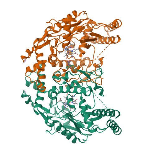
    Three nitric oxide synthase (NOS) isoforms, eNOS, nNOS and iNOS, generate nitric oxide (NO) crucial to the cardiovascular, nervous and host defense systems, respectively. Development of isoform-selective NOS inhibitors is of considerable therapeutic importance. Crystal structures of nNOS-selective dipeptide inhibitors in complex with both nNOS and eNOS were solved and the inhibitors were found to adopt a curled conformation in nNOS but an extended conformation in eNOS. We hypothesized that a single-residue difference in the active site, Asp597 (nNOS) versus Asn368 (eNOS), is responsible for the favored binding in nNOS. In the D597N nNOS mutant crystal structure, a bound inhibitor switches to the extended conformation and its inhibition of nNOS decreases >200-fold. Therefore, a single-residue difference is responsible for more than two orders of magnitude selectivity in inhibition of nNOS over eNOS by L-N(omega)-nitroarginine-containing dipeptide inhibitors.


  • Organizational Affiliation

    Department of Molecular Biology and Biochemistry and the Program in Macromolecular Structure, University of California, Irvine, California 92697-3900, USA.


Macromolecules
Find similar proteins by:  (by identity cutoff)  |  3D Structure
Entity ID: 1
MoleculeChains Sequence LengthOrganismDetailsImage
Nitric-oxide synthase, endothelial
A, B
417Bos taurusMutation(s): 0 
Gene Names: endothelial nitric oxide synthase
EC: 1.14.13.39
UniProt
Find proteins for P29473 (Bos taurus)
Explore P29473 
Go to UniProtKB:  P29473
Entity Groups  
Sequence Clusters30% Identity50% Identity70% Identity90% Identity95% Identity100% Identity
UniProt GroupP29473
Sequence Annotations
Expand
  • Reference Sequence
Small Molecules
Ligands 7 Unique
IDChains Name / Formula / InChI Key2D Diagram3D Interactions
HEM
Query on HEM

Download Ideal Coordinates CCD File 
E [auth A],
L [auth B]
PROTOPORPHYRIN IX CONTAINING FE
C34 H32 Fe N4 O4
KABFMIBPWCXCRK-RGGAHWMASA-L
DP3
Query on DP3

Download Ideal Coordinates CCD File 
G [auth A],
N [auth B]
N-{(4S)-4-AMINO-5-[(2-AMINOETHYL)AMINO]PENTYL}-N'-NITROGUANIDINE
C8 H21 N7 O2
RMSWBHUVFNFNIZ-ZETCQYMHSA-N
H4B
Query on H4B

Download Ideal Coordinates CCD File 
F [auth A],
M [auth B]
5,6,7,8-TETRAHYDROBIOPTERIN
C9 H15 N5 O3
FNKQXYHWGSIFBK-RPDRRWSUSA-N
CAC
Query on CAC

Download Ideal Coordinates CCD File 
C [auth A],
I [auth B]
CACODYLATE ION
C2 H6 As O2
OGGXGZAMXPVRFZ-UHFFFAOYSA-M
GOL
Query on GOL

Download Ideal Coordinates CCD File 
H [auth A],
O [auth B]
GLYCEROL
C3 H8 O3
PEDCQBHIVMGVHV-UHFFFAOYSA-N
ZN
Query on ZN

Download Ideal Coordinates CCD File 
K [auth B]ZINC ION
Zn
PTFCDOFLOPIGGS-UHFFFAOYSA-N
ACT
Query on ACT

Download Ideal Coordinates CCD File 
D [auth A],
J [auth B]
ACETATE ION
C2 H3 O2
QTBSBXVTEAMEQO-UHFFFAOYSA-M
Experimental Data & Validation

Experimental Data

  • Method: X-RAY DIFFRACTION
  • Resolution: 2.27 Å
  • R-Value Free:  0.278 (Depositor), 0.270 (DCC) 
  • R-Value Work:  0.215 (Depositor), 0.200 (DCC) 
  • R-Value Observed: 0.215 (Depositor) 
Space Group: P 21 21 21
Unit Cell:
Length ( Å )Angle ( ˚ )
a = 58.033α = 90
b = 106.085β = 90
c = 156.667γ = 90
Software Package:
Software NamePurpose
CNSrefinement
HKL-2000data reduction
SCALEPACKdata scaling
CNSphasing

Structure Validation

View Full Validation Report


Ligand Structure Quality Assessment 

Created with Raphaël 2.3.0Worse 01 BetterLigand structure goodness of fit to experimental dataBest fitted HEMClick on this verticalbar to view detailsBest fitted DP3Click on this verticalbar to view detailsBest fitted H4BClick on this verticalbar to view details

Entry History 

Deposition Data

Revision History  (Full details and data files)

  • Version 1.0: 2004-01-13
    Type: Initial release
  • Version 1.1: 2008-04-29
    Changes: Version format compliance
  • Version 1.2: 2011-07-13
    Changes: Non-polymer description, Version format compliance
  • Version 1.3: 2024-02-14
    Changes: Data collection, Database references, Derived calculations