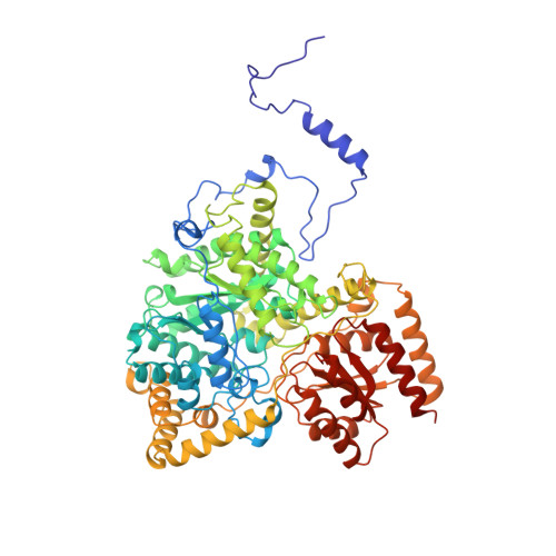
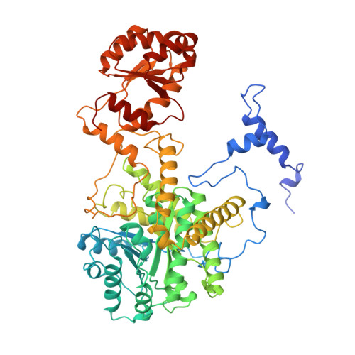
1REQ

METHYLMALONYL-COA MUTASE


Experimental Data Snapshot

  • Method: X-RAY DIFFRACTION
  • Resolution: 2.00 Å
  • R-Value Free: 0.275 
  • R-Value Work: 0.220 

wwPDB Validation   3D Report Full Report


Ligand Structure Quality Assessment 


This is version 1.4 of the entry. See complete history


Literature

How coenzyme B12 radicals are generated: the crystal structure of methylmalonyl-coenzyme A mutase at 2 A resolution.

Mancia, F.Keep, N.H.Nakagawa, A.Leadlay, P.F.McSweeney, S.Rasmussen, B.Bosecke, P.Diat, O.Evans, P.R.

(1996) Structure 4: 339-350

  • DOI: https://doi.org/10.1016/s0969-2126(96)00037-8
  • Primary Citation of Related Structures:  
    1REQ

  • PubMed Abstract: 

    The enzyme methylmalonyl-coenzyme A (CoA) mutase, an alphabeta heterodimer of 150 kDa, is a member of a class of enzymes that uses coenzyme B12 (adenosylcobalamin) as a cofactor. The enzyme induces the formation of an adenosyl radical from the cofactor. This radical then initiates a free-radical rearrangement of its substrate, succinyl-CoA, to methylmalonyl-CoA. Reported here is the crystal structure at 2 A resolution of methylmalonyl-CoA mutase from Propionibacterium shermanii in complex with coenzyme B12 and with the partial substrate desulpho-CoA (lacking the succinyl group and the sulphur atom of the substrate). The coenzyme is bound by a domain which shares a similar fold to those of flavodoxin and the B12-binding domain of methylcobalamin-dependent methionine synthase. The cobalt atom is coordinated, via a long bond, to a histidine from the protein. The partial substrate is bound along the axis of a (beta/alpha)8 TIM barrel domain. The histidine-cobalt distance is very long (2.5 A compared with 1.95-2.2 A in free cobalamins), suggesting that the enzyme positions the histidine in order to weaken the metal-carbon bond of the cofactor and favour the formation of the initial radical species. The active site is deeply buried, and the only access to it is through a narrow tunnel along the axis of the TIM barrel domain.


  • Organizational Affiliation

    MRC Laboratory of Molecular Biology, Hills Road, Cambridge CB2 2QH, UK.


Macromolecules
Find similar proteins by:  (by identity cutoff)  |  3D Structure
Entity ID: 1
MoleculeChains Sequence LengthOrganismDetailsImage
METHYLMALONYL-COA MUTASE
A, C
727Propionibacterium freudenreichii subsp. shermaniiMutation(s): 0 
Gene Names: MUTA MUTB
EC: 5.4.99.2
UniProt
Find proteins for P11653 (Propionibacterium freudenreichii subsp. shermanii)
Explore P11653 
Go to UniProtKB:  P11653
Entity Groups  
Sequence Clusters30% Identity50% Identity70% Identity90% Identity95% Identity100% Identity
UniProt GroupP11653
Sequence Annotations
Expand
  • Reference Sequence
Find similar proteins by:  (by identity cutoff)  |  3D Structure
Entity ID: 2
MoleculeChains Sequence LengthOrganismDetailsImage
METHYLMALONYL-COA MUTASE
B, D
637Propionibacterium freudenreichii subsp. shermaniiMutation(s): 0 
Gene Names: MUTA MUTB
EC: 5.4.99.2
UniProt
Find proteins for P11652 (Propionibacterium freudenreichii subsp. shermanii)
Explore P11652 
Go to UniProtKB:  P11652
Entity Groups  
Sequence Clusters30% Identity50% Identity70% Identity90% Identity95% Identity100% Identity
UniProt GroupP11652
Sequence Annotations
Expand
  • Reference Sequence
Small Molecules
Ligands 3 Unique
IDChains Name / Formula / InChI Key2D Diagram3D Interactions
B12
Query on B12

Download Ideal Coordinates CCD File 
E [auth A],
L [auth C]
COBALAMIN
C62 H89 Co N13 O14 P
LKVIQTCSMMVGFU-DWSMJLPVSA-N
DCA
Query on DCA

Download Ideal Coordinates CCD File 
F [auth A],
M [auth C]
DESULFO-COENZYME A
C21 H36 N7 O16 P3
ILWZMFJBPIYQKW-IBOSZNHHSA-N
GOL
Query on GOL

Download Ideal Coordinates CCD File 
G [auth A]
H [auth A]
I [auth B]
J [auth B]
K [auth B]
G [auth A],
H [auth A],
I [auth B],
J [auth B],
K [auth B],
N [auth C],
O [auth C],
P [auth D],
Q [auth D]
GLYCEROL
C3 H8 O3
PEDCQBHIVMGVHV-UHFFFAOYSA-N
Experimental Data & Validation

Experimental Data

  • Method: X-RAY DIFFRACTION
  • Resolution: 2.00 Å
  • R-Value Free: 0.275 
  • R-Value Work: 0.220 
  • Space Group: P 1 21 1
Unit Cell:
Length ( Å )Angle ( ˚ )
a = 119.8α = 90
b = 161.3β = 105.1
c = 88.4γ = 90
Software Package:
Software NamePurpose
REFMACrefinement
MOSFLMdata reduction

Structure Validation

View Full Validation Report


Ligand Structure Quality Assessment 


Entry History 

Deposition Data

Revision History  (Full details and data files)

  • Version 1.0: 1997-01-27
    Type: Initial release
  • Version 1.1: 2008-03-24
    Changes: Version format compliance
  • Version 1.2: 2011-07-13
    Changes: Non-polymer description, Version format compliance
  • Version 1.3: 2012-10-24
    Changes: Non-polymer description
  • Version 1.4: 2024-02-14
    Changes: Data collection, Database references, Derived calculations, Other