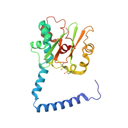
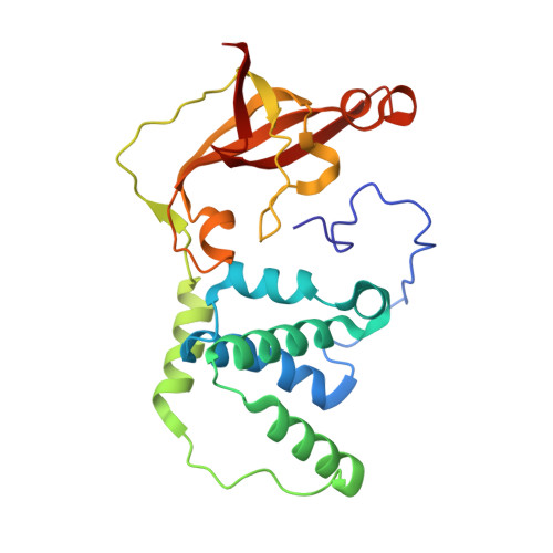
1UGR

Crystal structure of aT109S mutant of Co-type nitrile hydratase


Experimental Data Snapshot

  • Method: X-RAY DIFFRACTION
  • Resolution: 1.80 Å
  • R-Value Free: 0.213 
  • R-Value Work: 0.189 
  • R-Value Observed: 0.189 

wwPDB Validation   3D Report Full Report


This is version 1.4 of the entry. See complete history


Literature

Mutational and structural analysis of cobalt-containing nitrile hydratase on substrate and metal binding

Miyanaga, A.Fushinobu, S.Ito, K.Shoun, H.Wakagi, T.

(2004) Eur J Biochem 271: 429-438

  • DOI: https://doi.org/10.1046/j.1432-1033.2003.03943.x
  • Primary Citation of Related Structures:  
    1UGP, 1UGQ, 1UGR, 1UGS

  • PubMed Abstract: 

    Mutants of a cobalt-containing nitrile hydratase (NHase, EC 4.2.1.84) from Pseudonocardia thermophila JCM 3095 involved in substrate binding, catalysis and formation of the active center were constructed, and their characteristics and crystal structures were investigated. As expected from the structure of the substrate binding pocket, the wild-type enzyme showed significantly lower K(m) and K(i) values for aromatic substrates and inhibitors, respectively, than aliphatic ones. In the crystal structure of a complex with an inhibitor (n-butyric acid) the hydroxyl group of betaTyr68 formed hydrogen bonds with both n-butyric acid and alphaSer112, which is located in the active center. The betaY68F mutant showed an elevated K(m) value and a significantly decreased k(cat) value. The apoenzyme, which contains no detectable cobalt atom, was prepared from Escherichia coli cells grown in medium without cobalt ions. It showed no detectable activity. A disulfide bond between alphaCys108 and alphaCys113 was formed in the apoenzyme structure. In the highly conserved sequence motif in the cysteine cluster region, two positions are exclusively conserved in cobalt-containing or iron-containing nitrile hydratases. Two mutants (alphaT109S and alphaY114T) were constructed, each residue being replaced with an iron-containing one. The alphaT109S mutant showed similar characteristics to the wild-type enzyme. However, the alphaY114T mutant showed a very low cobalt content and catalytic activity compared with the wild-type enzyme, and oxidative modifications of alphaCys111 and alphaCys113 residues were not observed. The alphaTyr114 residue may be involved in the interaction with the nitrile hydratase activator protein of P. thermophila.


  • Organizational Affiliation

    Department of Biotechnology, The University of Tokyo, Japan.


Macromolecules
Find similar proteins by:  (by identity cutoff)  |  3D Structure
Entity ID: 1
MoleculeChains Sequence LengthOrganismDetailsImage
Nitrile Hydratase alpha subunit203Pseudonocardia thermophilaMutation(s): 3 
EC: 4.2.1.84
UniProt
Find proteins for Q7SID2 (Pseudonocardia thermophila)
Explore Q7SID2 
Go to UniProtKB:  Q7SID2
Entity Groups  
Sequence Clusters30% Identity50% Identity70% Identity90% Identity95% Identity100% Identity
UniProt GroupQ7SID2
Sequence Annotations
Expand
  • Reference Sequence
Find similar proteins by:  (by identity cutoff)  |  3D Structure
Entity ID: 2
MoleculeChains Sequence LengthOrganismDetailsImage
Nitrile Hydratase beta subunit228Pseudonocardia thermophilaMutation(s): 0 
EC: 4.2.1.84
UniProt
Find proteins for Q7SID3 (Pseudonocardia thermophila)
Explore Q7SID3 
Go to UniProtKB:  Q7SID3
Entity Groups  
Sequence Clusters30% Identity50% Identity70% Identity90% Identity95% Identity100% Identity
UniProt GroupQ7SID3
Sequence Annotations
Expand
  • Reference Sequence
Small Molecules
Ligands 1 Unique
IDChains Name / Formula / InChI Key2D Diagram3D Interactions
CO
Query on CO

Download Ideal Coordinates CCD File 
C [auth A]COBALT (II) ION
Co
XLJKHNWPARRRJB-UHFFFAOYSA-N
Modified Residues  2 Unique
IDChains TypeFormula2D DiagramParent
CSD
Query on CSD
A
L-PEPTIDE LINKINGC3 H7 N O4 SCYS
CSO
Query on CSO
A
L-PEPTIDE LINKINGC3 H7 N O3 SCYS
Experimental Data & Validation

Experimental Data

  • Method: X-RAY DIFFRACTION
  • Resolution: 1.80 Å
  • R-Value Free: 0.213 
  • R-Value Work: 0.189 
  • R-Value Observed: 0.189 
  • Space Group: P 32 2 1
Unit Cell:
Length ( Å )Angle ( ˚ )
a = 65.437α = 90
b = 65.437β = 90
c = 184.257γ = 120
Software Package:
Software NamePurpose
CNSrefinement
HKL-2000data reduction
SCALEPACKdata scaling
CNSphasing

Structure Validation

View Full Validation Report


Entry History 

Deposition Data

Revision History  (Full details and data files)

  • Version 1.0: 2004-06-17
    Type: Initial release
  • Version 1.1: 2008-04-27
    Changes: Version format compliance
  • Version 1.2: 2011-07-13
    Changes: Version format compliance
  • Version 1.3: 2021-11-10
    Changes: Database references, Derived calculations
  • Version 1.4: 2023-12-27
    Changes: Data collection