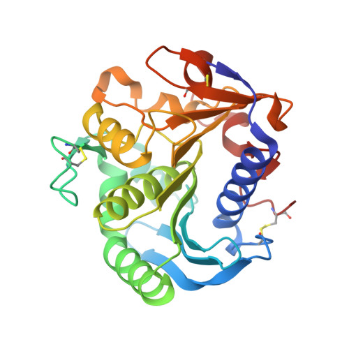
1UZA

Crystallographic structure of a feruloyl esterase from Aspergillus niger


Experimental Data Snapshot

  • Method: X-RAY DIFFRACTION
  • Resolution: 1.50 Å
  • R-Value Free: 0.188 
  • R-Value Work: 0.156 
  • R-Value Observed: 0.158 

wwPDB Validation   3D Report Full Report


Ligand Structure Quality Assessment 


This is version 1.2 of the entry. See complete history


Literature

Structure of a Feruloyl Esterase from Aspergillus Niger

Mcauley, K.E.Svendsen, A.Patkar, S.A.Wilson, K.S.

(2004) Acta Crystallogr D Biol Crystallogr 60: 878

  • DOI: https://doi.org/10.1107/S0907444904004937
  • Primary Citation of Related Structures:  
    1UWC, 1UZA

  • PubMed Abstract: 
  • The crystallographic structure of feruloyl esterase from Aspergillus niger has been determined to a resolution of 1.5 A by molecular replacement. The protein has an alpha/beta-hydrolase structure with a Ser-His-Asp catalytic triad; the overall fold of the protein is very similar to that of the fungal lipases ...

    The crystallographic structure of feruloyl esterase from Aspergillus niger has been determined to a resolution of 1.5 A by molecular replacement. The protein has an alpha/beta-hydrolase structure with a Ser-His-Asp catalytic triad; the overall fold of the protein is very similar to that of the fungal lipases. The structure of the enzyme-product complex was determined to a resolution of 1.08 A and reveals dual conformations for the serine and histidine residues at the active site.


    Organizational Affiliation

    CCLRC Daresbury Laboratory, Warrington, Cheshire WA4 4AD, England.


Macromolecules
Find similar proteins by:  (by identity cutoff)  |  3D Structure
Entity ID: 1
MoleculeChainsSequence LengthOrganismDetailsImage
FERULOYL ESTERASE A
A, B
261Aspergillus nigerMutation(s): 0 
Gene Names: faeA
EC: 3.1.1.73
UniProt
Find proteins for O42807 (Aspergillus niger)
Explore O42807 
Go to UniProtKB:  O42807
Entity Groups  
Sequence Clusters30% Identity50% Identity70% Identity90% Identity95% Identity100% Identity
UniProt GroupO42807
Protein Feature View
Expand
  • Reference Sequence
Small Molecules
Ligands 2 Unique
IDChainsName / Formula / InChI Key2D Diagram3D Interactions
NAG
Query on NAG

Download Ideal Coordinates CCD File 
C [auth A],
E [auth B]
2-acetamido-2-deoxy-beta-D-glucopyranose
C8 H15 N O6
OVRNDRQMDRJTHS-FMDGEEDCSA-N
 Ligand Interaction
SO4
Query on SO4

Download Ideal Coordinates CCD File 
D [auth A],
F [auth B]
SULFATE ION
O4 S
QAOWNCQODCNURD-UHFFFAOYSA-L
 Ligand Interaction
Experimental Data & Validation

Experimental Data

  • Method: X-RAY DIFFRACTION
  • Resolution: 1.50 Å
  • R-Value Free: 0.188 
  • R-Value Work: 0.156 
  • R-Value Observed: 0.158 
  • Space Group: P 1
Unit Cell:
Length ( Å )Angle ( ˚ )
a = 38.298α = 75.24
b = 39.681β = 78.82
c = 77.071γ = 71.3
Software Package:
Software NamePurpose
REFMACrefinement
DENZOdata reduction
SCALEPACKdata scaling
AMoREphasing

Structure Validation

View Full Validation Report


Ligand Structure Quality Assessment 


Entry History 

Deposition Data

Revision History  (Full details and data files)

  • Version 1.0: 2004-10-19
    Type: Initial release
  • Version 1.1: 2012-11-14
    Changes: Database references, Derived calculations, Non-polymer description, Other, Version format compliance
  • Version 1.2: 2020-07-29
    Type: Remediation
    Reason: Carbohydrate remediation
    Changes: Data collection, Derived calculations, Other, Structure summary