1WMQ | pdb_00001wmq

Structure of the HutP antitermination complex bound to a single stranded region of hut mRNA


Experimental Data Snapshot

  • Method: X-RAY DIFFRACTION
  • Resolution: 1.60 Å
  • R-Value Free: 
    0.249 (Depositor), 0.230 (DCC) 
  • R-Value Work: 
    0.225 (Depositor), 0.230 (DCC) 
  • R-Value Observed: 
    0.225 (Depositor) 

Starting Model: experimental
View more details

wwPDB Validation   3D Report Full Report


Ligand Structure Quality Assessment 

Created with Raphaël 2.3.0Worse 01 BetterLigand structure goodness of fit to experimental dataBest fitted HISClick on this verticalbar to view details

This is version 1.5 of the entry. See complete history


Literature

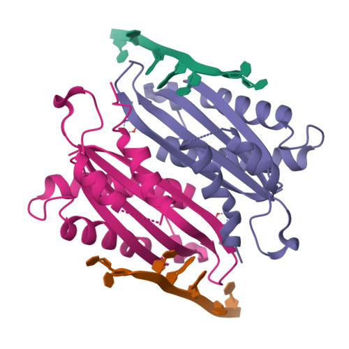
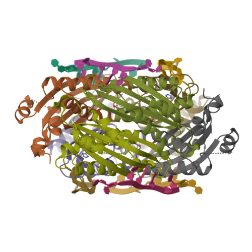
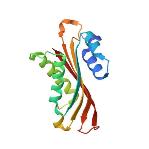
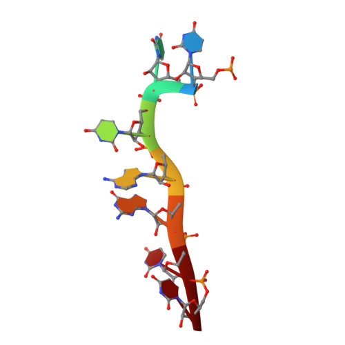
Structural basis of HutP-mediated anti-termination and roles of the Mg2+ ion and L-histidine ligand.

Kumarevel, T.Mizuno, H.Kumar, P.K.

(2005) Nature 434: 183-191

  • DOI: https://doi.org/10.1038/nature03355
  • Primary Citation of Related Structures:  
    1WMQ, 1WPS, 1WPV

  • PubMed Abstract: 

    HutP regulates the expression of the hut structural genes of Bacillus subtilis by an anti-termination mechanism and requires two components, Mg2+ ions and L-histidine. HutP recognizes three UAG triplet units, separated by four non-conserved nucleotides on the terminator region. Here we report the 1.60-A resolution crystal structure of the quaternary complex (HutP-L-histidine-Mg2+-21-base single-stranded RNA). In the complex, the RNA adopts a novel triangular fold on the hexameric surface of HutP, without any base-pairing, and binds to the protein mostly by specific protein-base interactions. The structure explains how the HutP and RNA interactions are regulated critically by the l-histidine and Mg2+ ion through the structural rearrangement. To gain insights into these structural rearrangements, we solved two additional crystal structures (uncomplexed HutP and HutP-L-histidine-Mg2+) that revealed the intermediate structures of HutP (before forming an active structure) and the importance of the Mg2+ ion interactions in the complexes.


  • Organizational Affiliation

    Functional Nucleic Acids Group, Institute for Biological Resources and Functions, National Institute of Advanced Industrial Science and Technology (AIST), Central 6, 1-1-1 Higashi, Tsukuba, Ibaraki 305-8566, Japan.


Macromolecules

Find similar proteins by:  (by identity cutoff)  |  3D Structure
Entity ID: 2
MoleculeChains Sequence LengthOrganismDetailsImage
Hut operon positive regulatory proteinC [auth A],
D [auth B]
147Bacillus subtilisMutation(s): 1 
UniProt
Find proteins for P10943 (Bacillus subtilis (strain 168))
Explore P10943 
Go to UniProtKB:  P10943
Entity Groups  
Sequence Clusters30% Identity50% Identity70% Identity90% Identity95% Identity100% Identity
UniProt GroupP10943
Sequence Annotations
Expand
  • Reference Sequence
Experimental Data & Validation

Experimental Data

  • Method: X-RAY DIFFRACTION
  • Resolution: 1.60 Å
  • R-Value Free:  0.249 (Depositor), 0.230 (DCC) 
  • R-Value Work:  0.225 (Depositor), 0.230 (DCC) 
  • R-Value Observed: 0.225 (Depositor) 
Space Group: H 3
Unit Cell:
Length ( Å )Angle ( ˚ )
a = 76.21α = 90
b = 76.21β = 90
c = 133.56γ = 120
Software Package:
Software NamePurpose
CNSrefinement
HKL-2000data reduction
CCP4data scaling
MOLREPphasing
PHASERphasing

Structure Validation

View Full Validation Report


Ligand Structure Quality Assessment 

Created with Raphaël 2.3.0Worse 01 BetterLigand structure goodness of fit to experimental dataBest fitted HISClick on this verticalbar to view details

Entry History 

Deposition Data

Revision History  (Full details and data files)

  • Version 1.0: 2005-03-15
    Type: Initial release
  • Version 1.1: 2008-04-30
    Changes: Version format compliance
  • Version 1.2: 2011-07-13
    Changes: Version format compliance
  • Version 1.3: 2017-10-11
    Changes: Advisory, Refinement description
  • Version 1.4: 2021-11-10
    Changes: Advisory, Database references, Derived calculations
  • Version 1.5: 2023-10-25
    Changes: Data collection, Refinement description