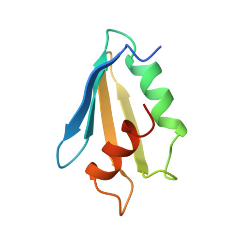
1XXA

C-TERMINAL DOMAIN OF ESCHERICHIA COLI ARGININE REPRESSOR/ L-ARGININE COMPLEX; PB DERIVATIVE


Experimental Data Snapshot

  • Method: X-RAY DIFFRACTION
  • Resolution: 2.20 Å
  • R-Value Free: 0.330 
  • R-Value Work: 0.200 
  • R-Value Observed: 0.200 

wwPDB Validation   3D Report Full Report


This is version 1.3 of the entry. See complete history


Literature

Structure of the oligomerization and L-arginine binding domain of the arginine repressor of Escherichia coli.

Van Duyne, G.D.Ghosh, G.Maas, W.K.Sigler, P.B.

(1996) J Mol Biol 256: 377-391

  • DOI: https://doi.org/10.1006/jmbi.1996.0093
  • Primary Citation of Related Structures:  
    1XXA, 1XXB, 1XXC

  • PubMed Abstract: 

    The structure of the oligomerization and L-arginine binding domain of the Escherichia coli arginine repressor (ArgR) has been determined using X-ray diffraction methods at 2.2 A resolution with bound arginine and at 2.8 A in the unliganded form. The oligomeric core is a 3-fold rotationally symmetric hexamer formed from six identical subunits corresponding to the 77 C-terminal residues (80 to 156) of ArgR. Each subunit has an alpha/beta fold containing a four-stranded antiparallel beta-sheet and two antiparallel alpha-helices. The hexamer is formed from two trimers, each with tightly packed hydrophobic cores. In the absence of arginine, the trimers stack back-to-back through a dyad-symmetric, sparsely packed hydrophobic interface. Six molecules of arginine bind at the trimer-trimer interface, each making ten hydrogen bonds to the protein including a direct ion pair that crosslinks the two protein trimers. Solution experiments with wild-type ArgR and oligomerization domain indicate that the hexameric form is greatly stabilized upon arginine binding. The crystal structures and solution experiments together suggest possible mechanisms of how arginine activates ArgR to bind to its DNA targets and provides a stereochemical basis for interpreting the results of mutagenesis and biochemical experiments with ArgR.


  • Organizational Affiliation

    Department of Molecular Biochemistry and Biophysics, Howard Hughes Medical Institute, Yale University, New Haven, CT 06510 USA.


Macromolecules
Find similar proteins by:  (by identity cutoff)  |  3D Structure
Entity ID: 1
MoleculeChains Sequence LengthOrganismDetailsImage
ARGININE REPRESSOR
A, B, C, D, E
A, B, C, D, E, F
78Escherichia coli K-12Mutation(s): 0 
Gene Names: T7
UniProt
Find proteins for P0A6D0 (Escherichia coli (strain K12))
Explore P0A6D0 
Go to UniProtKB:  P0A6D0
Entity Groups  
Sequence Clusters30% Identity50% Identity70% Identity90% Identity95% Identity100% Identity
UniProt GroupP0A6D0
Sequence Annotations
Expand
  • Reference Sequence
Small Molecules
Ligands 2 Unique
IDChains Name / Formula / InChI Key2D Diagram3D Interactions
PB
Query on PB

Download Ideal Coordinates CCD File 
I [auth C],
J [auth C],
L [auth D],
O [auth F]
LEAD (II) ION
Pb
RVPVRDXYQKGNMQ-UHFFFAOYSA-N
ARG
Query on ARG

Download Ideal Coordinates CCD File 
G [auth A]
H [auth A]
K [auth C]
M [auth D]
N [auth E]
G [auth A],
H [auth A],
K [auth C],
M [auth D],
N [auth E],
P [auth F]
ARGININE
C6 H15 N4 O2
ODKSFYDXXFIFQN-BYPYZUCNSA-O
Experimental Data & Validation

Experimental Data

  • Method: X-RAY DIFFRACTION
  • Resolution: 2.20 Å
  • R-Value Free: 0.330 
  • R-Value Work: 0.200 
  • R-Value Observed: 0.200 
  • Space Group: C 2 2 21
Unit Cell:
Length ( Å )Angle ( ˚ )
a = 53.5α = 90
b = 83.8β = 90
c = 217γ = 90
Software Package:
Software NamePurpose
DENZOdata reduction
SCALEPACKdata scaling
X-PLORmodel building
X-PLORrefinement
X-PLORphasing

Structure Validation

View Full Validation Report


Entry History 

Deposition Data

Revision History  (Full details and data files)

  • Version 1.0: 1996-03-08
    Type: Initial release
  • Version 1.1: 2008-03-24
    Changes: Version format compliance
  • Version 1.2: 2011-07-13
    Changes: Version format compliance
  • Version 1.3: 2024-02-14
    Changes: Data collection, Database references, Derived calculations