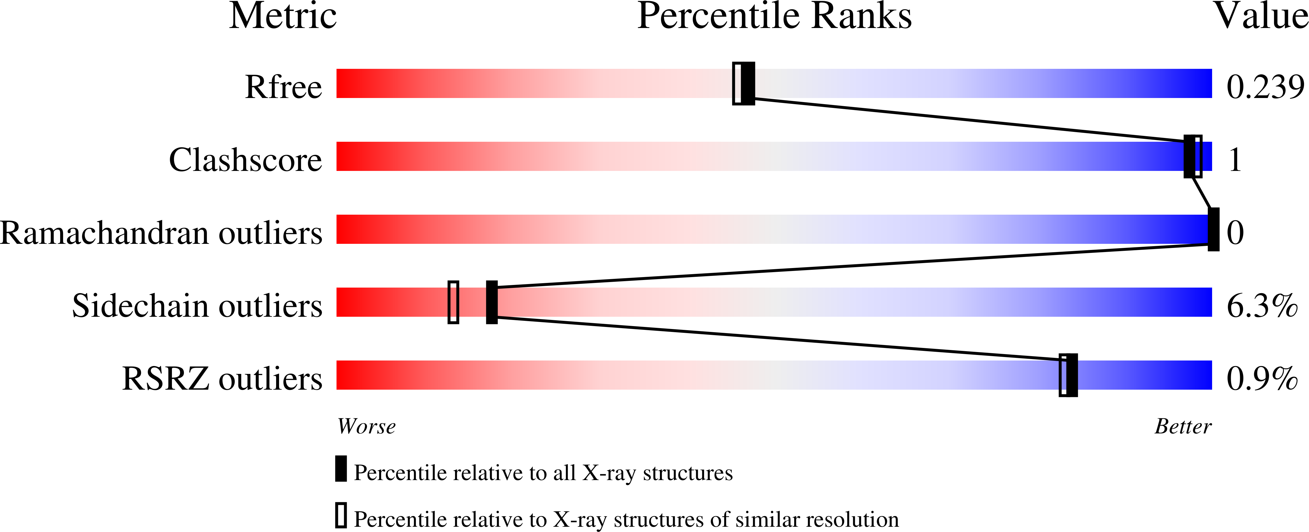
2AE0

Crystal structure of MltA from Escherichia coli reveals a unique lytic transglycosylase fold


Experimental Data Snapshot

  • Method: X-RAY DIFFRACTION
  • Resolution: 2.00 Å
  • R-Value Free: 0.237 
  • R-Value Work: 0.203 
  • R-Value Observed: 0.205 

wwPDB Validation   3D Report Full Report


This is version 1.4 of the entry. See complete history


Literature

Crystal Structure of MltA from Escherichia coli Reveals a Unique Lytic Transglycosylase Fold

van Straaten, K.E.Dijkstra, B.W.Vollmer, W.Thunnissen, A.M.W.H.

(2005) J Mol Biol 352: 1068-1080

  • DOI: https://doi.org/10.1016/j.jmb.2005.07.067
  • Primary Citation of Related Structures:  
    2AE0

  • PubMed Abstract: 

    Lytic transglycosylases are bacterial enzymes involved in the maintenance and growth of the bacterial cell-wall peptidoglycan. They cleave the beta-(1,4)-glycosidic bonds in peptidoglycan forming non-reducing 1,6-anhydromuropeptides. The crystal structure of the lytic transglycosylase MltA from Escherichia coli without a membrane anchor was solved at 2.0A resolution. The enzyme has a fold completely different from those of the other known lytic transglycosylases. It contains two domains, the largest of which has a double-psi beta-barrel fold, similar to that of endoglucanase V from Humicola insolens. The smaller domain also has a beta-barrel fold topology, which is weakly related to that of the RNA-binding domain of ribosomal proteins L25 and TL5. A large groove separates the two domains, which can accommodate a glycan strand, as shown by molecular modelling. Several conserved residues, one of which is in a position equivalent to that of the catalytic acid of the H.insolens endoglucanase, flank this putative substrate-binding groove. Mutation of this residue, Asp308, abolished all activity of the enzyme, supporting the direct participation of this residue in catalysis.


  • Organizational Affiliation

    Laboratory of Biophysical Chemistry, University of Groningen, Nijenborgh 4, 9747 AG Groningen, The Netherlands.


Macromolecules
Find similar proteins by:  (by identity cutoff)  |  3D Structure
Entity ID: 1
MoleculeChains Sequence LengthOrganismDetailsImage
Membrane-bound lytic murein transglycosylase AA [auth X]345Escherichia coliMutation(s): 0 
Gene Names: mlta
EC: 3.2.1
UniProt
Find proteins for P0A935 (Escherichia coli (strain K12))
Explore P0A935 
Go to UniProtKB:  P0A935
Entity Groups  
Sequence Clusters30% Identity50% Identity70% Identity90% Identity95% Identity100% Identity
UniProt GroupP0A935
Sequence Annotations
Expand
  • Reference Sequence
Experimental Data & Validation

Experimental Data

  • Method: X-RAY DIFFRACTION
  • Resolution: 2.00 Å
  • R-Value Free: 0.237 
  • R-Value Work: 0.203 
  • R-Value Observed: 0.205 
  • Space Group: P 31 2 1
Unit Cell:
Length ( Å )Angle ( ˚ )
a = 103.873α = 90
b = 103.873β = 90
c = 109.777γ = 120
Software Package:
Software NamePurpose
DENZOdata reduction
SCALEPACKdata scaling
REFMACrefinement
PDB_EXTRACTdata extraction
SnBphasing
SOLVEphasing
RESOLVEphasing

Structure Validation

View Full Validation Report


Entry History 

Deposition Data

Revision History  (Full details and data files)

  • Version 1.0: 2005-10-04
    Type: Initial release
  • Version 1.1: 2008-04-30
    Changes: Version format compliance
  • Version 1.2: 2011-07-13
    Changes: Advisory, Version format compliance
  • Version 1.3: 2017-10-11
    Changes: Refinement description
  • Version 1.4: 2024-03-13
    Changes: Data collection, Database references, Derived calculations