2AEP

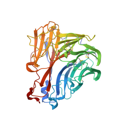
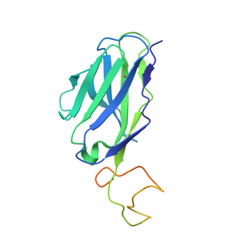
An epidemiologically significant epitope of a 1998 influenza virus neuraminidase forms a highly hydrated interface in the NA-antibody complex.


Experimental Data Snapshot

  • Method: X-RAY DIFFRACTION
  • Resolution: 2.10 Å
  • R-Value Free: 0.224 
  • R-Value Work: 0.188 
  • R-Value Observed: 0.190 

Starting Models: experimental
View more details

wwPDB Validation   3D Report Full Report


Ligand Structure Quality Assessment 


This is version 2.1 of the entry. See complete history


Literature

An Epidemiologically Significant Epitope of a 1998 Human Influenza Virus Neuraminidase Forms a Highly Hydrated Interface in the NA-Antibody Complex

Venkatramani, L.Bochkareva, E.Lee, J.T.Gulati, U.Laver, W.G.Bochkarev, A.Air, G.M.

(2006) J Mol Biol 356: 651-663

  • DOI: https://doi.org/10.1016/j.jmb.2005.11.061
  • Primary Citation of Related Structures:  
    2AEP, 2AEQ

  • PubMed Abstract: 

    The crystal structure of the complex between neuraminidase (NA) of influenza virus A/Memphis/31/98 (H3N2) and Fab of monoclonal antibody Mem5 has been determined at 2.1A resolution and shows a novel pattern of interactions compared to other NA-Fab structures. The interface buries a large area of 2400 A2 and the surfaces have high complementarity. However, the interface is also highly hydrated. There are 33 water molecules in the interface>or=95% buried from bulk solvent, but only 13 of these are isolated from other water molecules. The rest are involved in an intricate network of water-mediated hydrogen bonds throughout the interface, stabilizing the complex. Glu199 on NA, the most critical side-chain to the interaction as previously determined by escape mutant analysis and site-directed mutation, is located in a non-aqueous island. Glu199 and three other residues that contribute the major part of the antigen buried surface of the complex have mutated in human influenza viruses isolated after 1998, confirming that Mem5 identifies an epidemiologically important antigenic site. We conclude that antibody selection of NA variants is a significant component of recent antigenic drift in human H3N2 influenza viruses, supporting the idea that influenza vaccines should contain NA in addition to hemagglutinin.


  • Organizational Affiliation

    Department of Biochemistry and Molecular Biology, University of Oklahoma Health Sciences Center, Oklahoma City, OK 73190, USA.


Macromolecules
Find similar proteins by:  (by identity cutoff)  |  3D Structure
Entity ID: 1
MoleculeChains Sequence LengthOrganismDetailsImage
neuraminidase395Influenza A virus (A/Memphis/31/98(H3N2))Mutation(s): 0 
EC: 3.2.1.18
UniProt
Find proteins for Q9DL16 (Influenza A virus)
Explore Q9DL16 
Go to UniProtKB:  Q9DL16
Entity Groups  
Sequence Clusters30% Identity50% Identity70% Identity90% Identity95% Identity100% Identity
UniProt GroupQ9DL16
Glycosylation
Glycosylation Sites: 5
Sequence Annotations
Expand
  • Reference Sequence
Find similar proteins by:  (by identity cutoff)  |  3D Structure
Entity ID: 2
MoleculeChains Sequence LengthOrganismDetailsImage
FAB heavy chainB [auth H]217Mus musculusMutation(s): 0 
UniProt
Find proteins for P84751 (Mus musculus)
Explore P84751 
Go to UniProtKB:  P84751
Entity Groups  
Sequence Clusters30% Identity50% Identity70% Identity90% Identity95% Identity100% Identity
UniProt GroupP84751
Sequence Annotations
Expand
  • Reference Sequence
Find similar proteins by:  (by identity cutoff)  |  3D Structure
Entity ID: 3
MoleculeChains Sequence LengthOrganismDetailsImage
FAB light chainC [auth L]214Mus musculusMutation(s): 0 
UniProt
Find proteins for P84750 (Mus musculus)
Explore P84750 
Go to UniProtKB:  P84750
Entity Groups  
Sequence Clusters30% Identity50% Identity70% Identity90% Identity95% Identity100% Identity
UniProt GroupP84750
Sequence Annotations
Expand
  • Reference Sequence
Oligosaccharides

Help

Entity ID: 4
MoleculeChains Length2D Diagram Glycosylation3D Interactions
alpha-D-mannopyranose-(1-2)-alpha-D-mannopyranose-(1-3)-[alpha-D-mannopyranose-(1-6)-alpha-D-mannopyranose-(1-6)]beta-D-mannopyranose-(1-4)-2-acetamido-2-deoxy-beta-D-glucopyranose-(1-4)-2-acetamido-2-deoxy-beta-D-glucopyranoseD [auth B]7N-Glycosylation
Glycosylation Resources
GlyTouCan:  G23799GS
GlyCosmos:  G23799GS
GlyGen:  G23799GS
Small Molecules
Ligands 5 Unique
IDChains Name / Formula / InChI Key2D Diagram3D Interactions
NAG
Query on NAG

Download Ideal Coordinates CCD File 
F [auth A],
G [auth A],
H [auth A],
I [auth A]
2-acetamido-2-deoxy-beta-D-glucopyranose
C8 H15 N O6
OVRNDRQMDRJTHS-FMDGEEDCSA-N
GLC
Query on GLC

Download Ideal Coordinates CCD File 
E [auth A],
L [auth H]
alpha-D-glucopyranose
C6 H12 O6
WQZGKKKJIJFFOK-DVKNGEFBSA-N
MAN
Query on MAN

Download Ideal Coordinates CCD File 
M [auth L]alpha-D-mannopyranose
C6 H12 O6
WQZGKKKJIJFFOK-PQMKYFCFSA-N
SO4
Query on SO4

Download Ideal Coordinates CCD File 
K [auth A]SULFATE ION
O4 S
QAOWNCQODCNURD-UHFFFAOYSA-L
CA
Query on CA

Download Ideal Coordinates CCD File 
J [auth A]CALCIUM ION
Ca
BHPQYMZQTOCNFJ-UHFFFAOYSA-N
Experimental Data & Validation

Experimental Data

  • Method: X-RAY DIFFRACTION
  • Resolution: 2.10 Å
  • R-Value Free: 0.224 
  • R-Value Work: 0.188 
  • R-Value Observed: 0.190 
  • Space Group: P 4 21 2
Unit Cell:
Length ( Å )Angle ( ˚ )
a = 155.057α = 90
b = 155.057β = 90
c = 102.645γ = 90
Software Package:
Software NamePurpose
REFMACrefinement
DENZOdata reduction
SCALEPACKdata scaling
AMoREphasing

Structure Validation

View Full Validation Report


Ligand Structure Quality Assessment 


Entry History 

Deposition Data

Revision History  (Full details and data files)

  • Version 1.0: 2005-12-20
    Type: Initial release
  • Version 1.1: 2008-04-30
    Changes: Version format compliance
  • Version 1.2: 2011-07-13
    Changes: Non-polymer description, Version format compliance
  • Version 2.0: 2020-07-29
    Type: Remediation
    Reason: Carbohydrate remediation
    Changes: Advisory, Atomic model, Data collection, Derived calculations, Structure summary
  • Version 2.1: 2023-08-23
    Changes: Data collection, Database references, Refinement description, Structure summary