2BGI

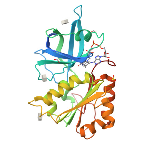
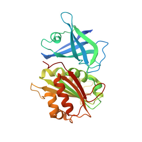
X-Ray Structure of the Ferredoxin-NADP(H) Reductase from Rhodobacter capsulatus complexed with three molecules of the detergent n-heptyl- beta-D-thioglucoside at 1.7 Angstroms


Experimental Data Snapshot

  • Method: X-RAY DIFFRACTION
  • Resolution: 1.68 Å
  • R-Value Free: 0.225 
  • R-Value Work: 0.221 
  • R-Value Observed: 0.221 

Starting Model: experimental
View more details

wwPDB Validation   3D Report Full Report


Ligand Structure Quality Assessment 


This is version 1.4 of the entry. See complete history


Literature

The Ferredoxin-Nadp(H) Reductase from Rhodobacter Capsulatus: Molecular Structure and Catalytic Mechanism

Nogues, I.Perez-Dorado, J.I.Frago, S.Bittel, C.Mayhew, S.G.Gomez-Moreno, C.Hermoso, J.A.Medina, M.Cortez, N.Carrillo, N.

(2005) Biochemistry 44: 11730

  • DOI: https://doi.org/10.1021/bi0508183
  • Primary Citation of Related Structures:  
    2BGI, 2BGJ

  • PubMed Abstract: 

    The photosynthetic bacterium Rhodobacter capsulatus contains a ferredoxin (flavodoxin)-NADP(H) oxidoreductase (FPR) that catalyzes electron transfer between NADP(H) and ferredoxin or flavodoxin. The structure of the enzyme, determined by X-ray crystallography, contains two domains harboring the FAD and NADP(H) binding sites, as is typical of the FPR structural family. The FAD molecule is in a hairpin conformation in which stacking interactions can be established between the dimethylisoalloxazine and adenine moieties. The midpoint redox potentials of the various transitions undergone by R. capsulatus FPR were similar to those reported for their counterparts involved in oxygenic photosynthesis, but its catalytic activity is orders of magnitude lower (1-2 s(-)(1) versus 200-500 s(-)(1)) as is true for most of its prokaryotic homologues. To identify the mechanistic basis for the slow turnover in the bacterial enzymes, we dissected the R. capsulatus FPR reaction into hydride transfer and electron transfer steps, and determined their rates using stopped-flow methods. Hydride exchange between the enzyme and NADP(H) occurred at 30-150 s(-)(1), indicating that this half-reaction does not limit FPR activity. In contrast, electron transfer to flavodoxin proceeds at 2.7 s(-)(1), in the range of steady-state catalysis. Flavodoxin semiquinone was a better electron acceptor for FPR than oxidized flavodoxin under both single turnover and steady-state conditions. The results indicate that one-electron reduction of oxidized flavodoxin limits the enzyme activity in vitro, and support the notion that flavodoxin oscillates between the semiquinone and fully reduced states when FPR operates in vivo.


  • Organizational Affiliation

    Departamento de Bioquímica y Biología Molecular y Celular, Facultad de Ciencias, and Institute of Biocomputation and Physics of Complex Systems (BIFI), Universidad de Zaragoza, Zaragoza, Spain.


Macromolecules
Find similar proteins by:  (by identity cutoff)  |  3D Structure
Entity ID: 1
MoleculeChains Sequence LengthOrganismDetailsImage
FERREDOXIN-NADP(H) REDUCTASE272Rhodobacter capsulatusMutation(s): 0 
EC: 1.18.1.2 (PDB Primary Data), 1.19.1.1 (UniProt)
UniProt
Find proteins for Q9L6V3 (Rhodobacter capsulatus)
Explore Q9L6V3 
Go to UniProtKB:  Q9L6V3
Entity Groups  
Sequence Clusters30% Identity50% Identity70% Identity90% Identity95% Identity100% Identity
UniProt GroupQ9L6V3
Sequence Annotations
Expand
  • Reference Sequence
Small Molecules
Ligands 4 Unique
IDChains Name / Formula / InChI Key2D Diagram3D Interactions
FAD
Query on FAD

Download Ideal Coordinates CCD File 
B [auth A]FLAVIN-ADENINE DINUCLEOTIDE
C27 H33 N9 O15 P2
VWWQXMAJTJZDQX-UYBVJOGSSA-N
HTG
Query on HTG

Download Ideal Coordinates CCD File 
C [auth A],
D [auth A],
F [auth A]
heptyl 1-thio-beta-D-glucopyranoside
C13 H26 O5 S
HPEGNLMTTNTJSP-LBELIVKGSA-N
SO4
Query on SO4

Download Ideal Coordinates CCD File 
E [auth A]SULFATE ION
O4 S
QAOWNCQODCNURD-UHFFFAOYSA-L
CO2
Query on CO2

Download Ideal Coordinates CCD File 
G [auth A],
H [auth A]
CARBON DIOXIDE
C O2
CURLTUGMZLYLDI-UHFFFAOYSA-N
Experimental Data & Validation

Experimental Data

  • Method: X-RAY DIFFRACTION
  • Resolution: 1.68 Å
  • R-Value Free: 0.225 
  • R-Value Work: 0.221 
  • R-Value Observed: 0.221 
  • Space Group: P 31 2 1
Unit Cell:
Length ( Å )Angle ( ˚ )
a = 120.848α = 90
b = 120.848β = 90
c = 50.709γ = 120
Software Package:
Software NamePurpose
CNSrefinement
MOSFLMdata reduction
SCALAdata scaling
AMoREphasing

Structure Validation

View Full Validation Report


Ligand Structure Quality Assessment 


Entry History 

Deposition Data

Revision History  (Full details and data files)

  • Version 1.0: 2005-09-07
    Type: Initial release
  • Version 1.1: 2011-05-08
    Changes: Version format compliance
  • Version 1.2: 2011-07-13
    Changes: Version format compliance
  • Version 1.3: 2020-07-29
    Type: Remediation
    Reason: Carbohydrate remediation
    Changes: Derived calculations, Other, Structure summary
  • Version 1.4: 2023-12-13
    Changes: Data collection, Database references, Refinement description, Structure summary