2BIG

Radiation damage of the Schiff base in phosphoserine aminotransferase (structure I)


Experimental Data Snapshot

  • Method: X-RAY DIFFRACTION
  • Resolution: 1.30 Å
  • R-Value Free: 0.156 
  • R-Value Observed: 0.115 

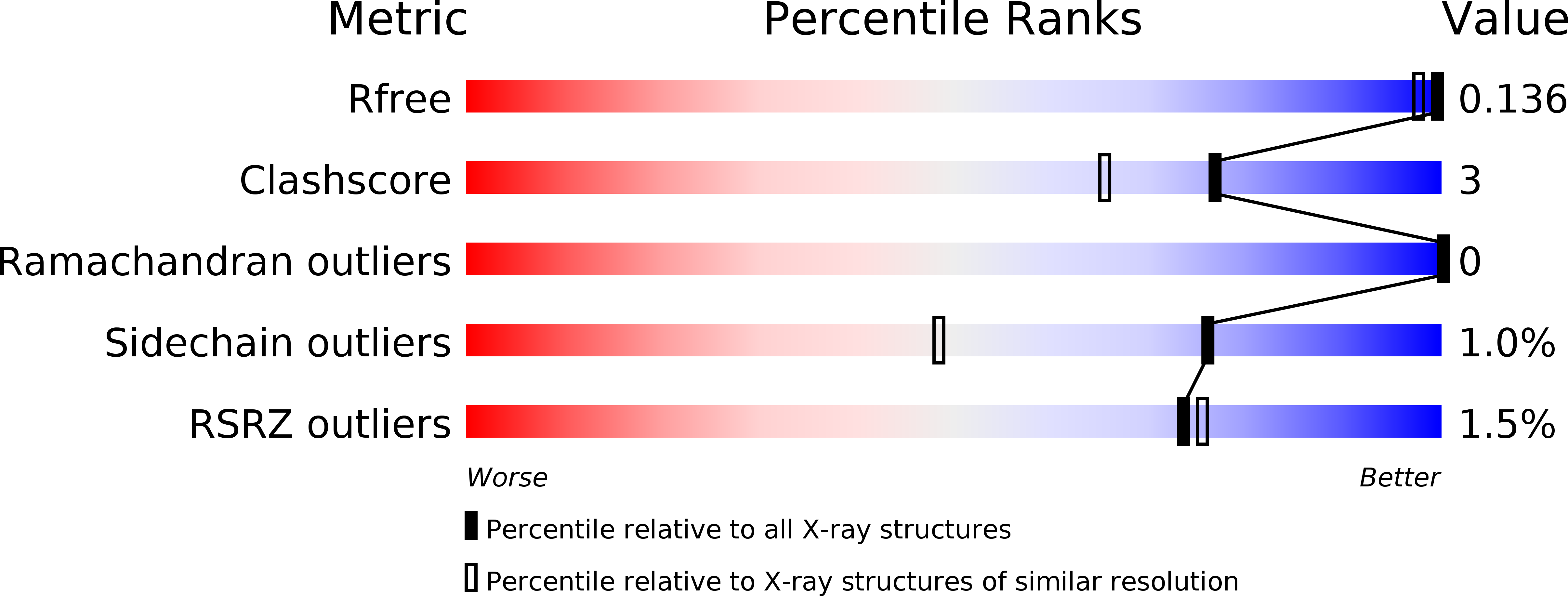
wwPDB Validation   3D Report Full Report


Ligand Structure Quality Assessment 


This is version 1.4 of the entry. See complete history


Literature

Strain Relief at the Active Site of Phosphoserine Aminotransferase Induced by Radiation Damage.

Dubnovitsky, A.P.Ravelli, R.B.G.Popov, A.N.Papageorgiou, A.C.

(2005) Protein Sci 14: 1498

  • DOI: https://doi.org/10.1110/ps.051397905
  • Primary Citation of Related Structures:  
    2BHX, 2BI1, 2BI2, 2BI3, 2BI5, 2BI9, 2BIA, 2BIE, 2BIG

  • PubMed Abstract: 

    The X-ray susceptibility of the lysine-pyridoxal-5'-phosphate Schiff base in Bacillus alcalophilus phosphoserine aminotransferase has been investigated using crystallographic data collected at 100 K to 1.3 A resolution, complemented by on-line spectroscopic studies. X-rays induce deprotonation of the internal aldimine, changes in the Schiff base conformation, displacement of the cofactor molecule, and disruption of the Schiff base linkage between pyridoxal-5'-phosphate and the Lys residue. Analysis of the "undamaged" structure reveals a significant chemical strain on the internal aldimine bond that leads to a pronounced geometrical distortion of the cofactor. However, upon crystal exposure to the X-rays, the strain and distortion are relaxed and eventually diminished when the total absorbed dose has exceeded 4.7 x 10(6) Ggamma. Our data provide new insights into the enzymatic activation of pyridoxal-5'-phosphate and suggest that special care should be taken while using macromolecular crystallography to study details in strained active sites.


  • Organizational Affiliation

    Turku Centre for Biotechnology, University of Turku, Finland.


Macromolecules
Find similar proteins by:  (by identity cutoff)  |  3D Structure
Entity ID: 1
MoleculeChains Sequence LengthOrganismDetailsImage
PHOSPHOSERINE AMINOTRANSFERASE
A, B
361Alkalihalobacillus alcalophilusMutation(s): 0 
EC: 2.6.1.52
UniProt
Find proteins for Q9RME2 (Alkalihalobacillus alcalophilus)
Explore Q9RME2 
Go to UniProtKB:  Q9RME2
Entity Groups  
Sequence Clusters30% Identity50% Identity70% Identity90% Identity95% Identity100% Identity
UniProt GroupQ9RME2
Sequence Annotations
Expand
  • Reference Sequence
Small Molecules
Ligands 5 Unique
IDChains Name / Formula / InChI Key2D Diagram3D Interactions
PLP
Query on PLP

Download Ideal Coordinates CCD File 
C [auth A],
J [auth B]
PYRIDOXAL-5'-PHOSPHATE
C8 H10 N O6 P
NGVDGCNFYWLIFO-UHFFFAOYSA-N
PGE
Query on PGE

Download Ideal Coordinates CCD File 
H [auth A]TRIETHYLENE GLYCOL
C6 H14 O4
ZIBGPFATKBEMQZ-UHFFFAOYSA-N
PEG
Query on PEG

Download Ideal Coordinates CCD File 
I [auth A],
P [auth B]
DI(HYDROXYETHYL)ETHER
C4 H10 O3
MTHSVFCYNBDYFN-UHFFFAOYSA-N
CL
Query on CL

Download Ideal Coordinates CCD File 
F [auth A],
G [auth A],
N [auth B],
O [auth B]
CHLORIDE ION
Cl
VEXZGXHMUGYJMC-UHFFFAOYSA-M
MG
Query on MG

Download Ideal Coordinates CCD File 
D [auth A],
E [auth A],
K [auth B],
L [auth B],
M [auth B]
MAGNESIUM ION
Mg
JLVVSXFLKOJNIY-UHFFFAOYSA-N
Experimental Data & Validation

Experimental Data

  • Method: X-RAY DIFFRACTION
  • Resolution: 1.30 Å
  • R-Value Free: 0.156 
  • R-Value Observed: 0.115 
  • Space Group: P 21 21 2
Unit Cell:
Length ( Å )Angle ( ˚ )
a = 143.801α = 90
b = 84.297β = 90
c = 67.264γ = 90
Software Package:
Software NamePurpose
SHELXL-97refinement
HKLdata reduction
HKLdata scaling

Structure Validation

View Full Validation Report


Ligand Structure Quality Assessment 


Entry History 

Deposition Data

Revision History  (Full details and data files)

  • Version 1.0: 2005-05-19
    Type: Initial release
  • Version 1.1: 2011-05-08
    Changes: Version format compliance
  • Version 1.2: 2011-07-13
    Changes: Version format compliance
  • Version 1.3: 2019-05-22
    Changes: Data collection, Derived calculations, Other, Refinement description
  • Version 1.4: 2019-07-24
    Changes: Data collection