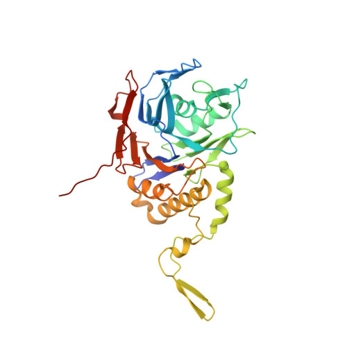
2BJF

Crystal Structure of Conjugated Bile Acid Hydrolase from Clostridium perfringens in Complex with Reaction Products Taurine and Deoxycholate


Experimental Data Snapshot

  • Method: X-RAY DIFFRACTION
  • Resolution: 1.67 Å
  • R-Value Free: 0.201 
  • R-Value Work: 0.182 
  • R-Value Observed: 0.183 

Starting Model: experimental
View more details

wwPDB Validation   3D Report Full Report


Ligand Structure Quality Assessment 


This is version 1.3 of the entry. See complete history


Literature

Conjugated Bile Acid Hydrolase is a Tetrameric N-Terminal Thiol Hydrolase with Specific Recognition of its Cholyl But not of its Tauryl Product

Rossocha, M.Schultz-Heienbrok, R.Von Moeller, H.Coleman, J.P.Saenger, W.

(2005) Biochemistry 44: 5739

  • DOI: https://doi.org/10.1021/bi0473206
  • Primary Citation of Related Structures:  
    2BJF, 2BJG

  • PubMed Abstract: 

    Bacterial bile salt hydrolases catalyze the degradation of conjugated bile acids in the mammalian gut. The crystal structures of conjugated bile acid hydrolase (CBAH) from Clostridium perfringens as apoenzyme and in complex with taurodeoxycholate that was hydrolyzed to the reaction products taurine and deoxycholate are described here at 2.1 and 1.7 A resolution, respectively. The crystal structures reveal close relationship between CBAH and penicillin V acylase from Bacillus sphaericus. This similarity together with the N-terminal cysteine classifies CBAH as a member of the N-terminal nucleophile (Ntn) hydrolase superfamily. Both crystal structures show an identical homotetrameric organization with dihedral (D(2) or 222) point group symmetry. The structure analysis of C. perfringens CBAH identifies critical residues in catalysis, substrate recognition, and tetramer formation which may serve in further biochemical characterization of bile acid hydrolases.


  • Organizational Affiliation

    Freie Universität Berlin, Institut für Kristallographie, Takustrasse 6, 14195 Berlin, Germany.


Macromolecules
Find similar proteins by:  (by identity cutoff)  |  3D Structure
Entity ID: 1
MoleculeChains Sequence LengthOrganismDetailsImage
CHOLOYLGLYCINE HYDROLASE329Clostridium perfringensMutation(s): 0 
EC: 3.5.1.24 (PDB Primary Data), 3.5.1 (UniProt), 2.3.1 (UniProt)
UniProt
Find proteins for P54965 (Clostridium perfringens (strain 13 / Type A))
Explore P54965 
Go to UniProtKB:  P54965
Entity Groups  
Sequence Clusters30% Identity50% Identity70% Identity90% Identity95% Identity100% Identity
UniProt GroupP54965
Sequence Annotations
Expand
  • Reference Sequence
Experimental Data & Validation

Experimental Data

  • Method: X-RAY DIFFRACTION
  • Resolution: 1.67 Å
  • R-Value Free: 0.201 
  • R-Value Work: 0.182 
  • R-Value Observed: 0.183 
  • Space Group: F 2 2 2
Unit Cell:
Length ( Å )Angle ( ˚ )
a = 88.848α = 90
b = 90.835β = 90
c = 188.547γ = 90
Software Package:
Software NamePurpose
REFMACrefinement
DENZOdata reduction
SCALEPACKdata scaling
MOLREPphasing

Structure Validation

View Full Validation Report


Ligand Structure Quality Assessment 


Entry History 

Deposition Data

Revision History  (Full details and data files)

  • Version 1.0: 2005-03-03
    Type: Initial release
  • Version 1.1: 2011-05-08
    Changes: Version format compliance
  • Version 1.2: 2011-07-13
    Changes: Version format compliance
  • Version 1.3: 2023-12-13
    Changes: Data collection, Database references, Derived calculations, Refinement description