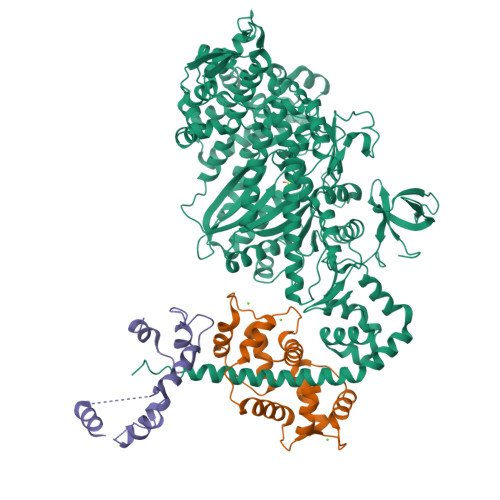
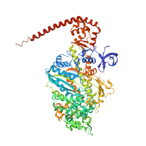
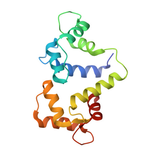
2BKI

Myosin VI nucleotide-free (MDinsert2-IQ) crystal structure


Experimental Data Snapshot

  • Method: X-RAY DIFFRACTION
  • Resolution: 2.90 Å
  • R-Value Free: 0.304 
  • R-Value Work: 0.264 
  • R-Value Observed: 0.266 

Starting Model: experimental
View more details

wwPDB Validation   3D Report Full Report


This is version 1.2 of the entry. See complete history


Literature

The Structure of the Myosin Vi Motor Reveals the Mechanism of Directionality Reversal

Menetrey, J.Bahloul, A.Wells, A.Yengo, C.Morris, C.Sweeney, H.L.Houdusse, A.

(2005) Nature 435: 779

  • DOI: https://doi.org/10.1038/nature03592
  • Primary Citation of Related Structures:  
    2BKH, 2BKI

  • PubMed Abstract: 

    Here we solve a 2.4-A structure of a truncated version of the reverse-direction myosin motor, myosin VI, that contains the motor domain and binding sites for two calmodulin molecules. The structure reveals only minor differences in the motor domain from that in plus-end directed myosins, with the exception of two unique inserts. The first is near the nucleotide-binding pocket and alters the rates of nucleotide association and dissociation. The second unique insert forms an integral part of the myosin VI converter domain along with a calmodulin bound to a novel target motif within the insert. This serves to redirect the effective 'lever arm' of myosin VI, which includes a second calmodulin bound to an 'IQ motif', towards the pointed (minus) end of the actin filament. This repositioning largely accounts for the reverse directionality of this class of myosin motors. We propose a model incorporating a kinesin-like uncoupling/docking mechanism to provide a full explanation of the movements of myosin VI.


  • Organizational Affiliation

    Structural Motility, Institut Curie CNRS, UMR144, 26 rue d'Ulm, 75248 Paris cedex 05, France.


Macromolecules
Find similar proteins by:  (by identity cutoff)  |  3D Structure
Entity ID: 1
MoleculeChains Sequence LengthOrganismDetailsImage
UNCONVENTIONAL MYOSIN858Sus scrofaMutation(s): 0 
UniProt
Find proteins for Q29122 (Sus scrofa)
Explore Q29122 
Go to UniProtKB:  Q29122
Entity Groups  
Sequence Clusters30% Identity50% Identity70% Identity90% Identity95% Identity100% Identity
UniProt GroupQ29122
Sequence Annotations
Expand
  • Reference Sequence
Find similar proteins by:  (by identity cutoff)  |  3D Structure
Entity ID: 2
MoleculeChains Sequence LengthOrganismDetailsImage
CALMODULINB,
C [auth D]
149Gallus gallusMutation(s): 0 
UniProt
Find proteins for P62149 (Gallus gallus)
Explore P62149 
Go to UniProtKB:  P62149
Entity Groups  
Sequence Clusters30% Identity50% Identity70% Identity90% Identity95% Identity100% Identity
UniProt GroupP62149
Sequence Annotations
Expand
  • Reference Sequence
Experimental Data & Validation

Experimental Data

  • Method: X-RAY DIFFRACTION
  • Resolution: 2.90 Å
  • R-Value Free: 0.304 
  • R-Value Work: 0.264 
  • R-Value Observed: 0.266 
  • Space Group: P 21 21 2
Unit Cell:
Length ( Å )Angle ( ˚ )
a = 104.781α = 90
b = 250.167β = 90
c = 67.274γ = 90
Software Package:
Software NamePurpose
REFMACrefinement
MOSFLMdata reduction
SCALAdata scaling
AMoREphasing

Structure Validation

View Full Validation Report


Entry History 

Deposition Data

Revision History  (Full details and data files)

  • Version 1.0: 2005-06-07
    Type: Initial release
  • Version 1.1: 2015-09-16
    Changes: Database references, Derived calculations, Non-polymer description, Other, Source and taxonomy, Structure summary, Version format compliance
  • Version 1.2: 2023-12-13
    Changes: Data collection, Database references, Derived calculations, Other, Refinement description