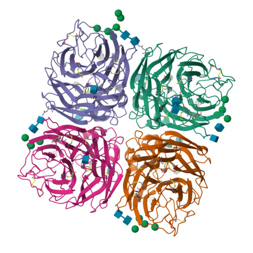
The Crystal Structure of Type a Influenza Virus Neuraminidase of the N6 Subtype Reveals the Existence of Two Separate Neu5Ac Binding Sites
Rudino-Pinera, E., Tunnah, P., Crennell, S.J., Webster, R.G., Laver, W.G., Garman, E.F.To be published.
Experimental Data Snapshot
Starting Model: experimental
View more details
Entity ID: 1 | |||||
|---|---|---|---|---|---|
| Molecule | Chains | Sequence Length | Organism | Details | Image |
| NEURAMINIDASE | 389 | Influenza A virus | Mutation(s): 0 EC: 3.2.1.18 | 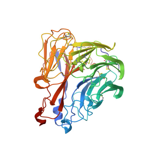 | |
UniProt | |||||
Find proteins for Q6XV27 (Influenza A virus (strain A/Duck/England/1/1956 H11N6)) Explore Q6XV27 Go to UniProtKB: Q6XV27 | |||||
Entity Groups | |||||
| Sequence Clusters | 30% Identity50% Identity70% Identity90% Identity95% Identity100% Identity | ||||
| UniProt Group | Q6XV27 | ||||
Glycosylation | |||||
| Glycosylation Sites: 3 | |||||
Sequence AnnotationsExpand | |||||
| |||||
Entity ID: 2 | |||||
|---|---|---|---|---|---|
| Molecule | Chains | Length | 2D Diagram | Glycosylation | 3D Interactions |
| 2-acetamido-2-deoxy-beta-D-glucopyranose-(1-4)-2-acetamido-2-deoxy-beta-D-glucopyranose | E, H, I | 2 |  | N-Glycosylation | |
Glycosylation Resources | |||||
GlyTouCan: G42666HT GlyCosmos: G42666HT GlyGen: G42666HT | |||||
Entity ID: 3 | |||||
|---|---|---|---|---|---|
| Molecule | Chains | Length | 2D Diagram | Glycosylation | 3D Interactions |
| alpha-D-mannopyranose-(1-3)-[alpha-D-mannopyranose-(1-6)]alpha-D-mannopyranose | F | 3 |  | N/A | |
Glycosylation Resources | |||||
GlyTouCan: G00619DD GlyCosmos: G00619DD GlyGen: G00619DD | |||||
Entity ID: 4 | |||||
|---|---|---|---|---|---|
| Molecule | Chains | Length | 2D Diagram | Glycosylation | 3D Interactions |
| alpha-D-mannopyranose-(1-3)-alpha-D-mannopyranose-(1-6)-beta-D-mannopyranose-(1-4)-2-acetamido-2-deoxy-beta-D-glucopyranose-(1-4)-2-acetamido-2-deoxy-beta-D-glucopyranose | G | 5 |  | N-Glycosylation | |
Glycosylation Resources | |||||
GlyTouCan: G10756ZZ GlyCosmos: G10756ZZ GlyGen: G10756ZZ | |||||
| Ligands 5 Unique | |||||
|---|---|---|---|---|---|
| ID | Chains | Name / Formula / InChI Key | 2D Diagram | 3D Interactions | |
| ZMR Query on ZMR | AA [auth C] J [auth A] JA [auth D] K [auth A] KA [auth D] | ZANAMIVIR C12 H20 N4 O7 ARAIBEBZBOPLMB-UFGQHTETSA-N |  | ||
| NAG Query on NAG | DA [auth C] EA [auth C] FA [auth C] M [auth A] N [auth A] | 2-acetamido-2-deoxy-beta-D-glucopyranose C8 H15 N O6 OVRNDRQMDRJTHS-FMDGEEDCSA-N |  | ||
| MAN Query on MAN | CA [auth C] GA [auth C] HA [auth C] MA [auth D] NA [auth D] | alpha-D-mannopyranose C6 H12 O6 WQZGKKKJIJFFOK-PQMKYFCFSA-N |  | ||
| BMA Query on BMA | IA [auth C], O [auth A], QA [auth D] | beta-D-mannopyranose C6 H12 O6 WQZGKKKJIJFFOK-RWOPYEJCSA-N |  | ||
| CA Query on CA | BA [auth C], L [auth A], LA [auth D], R [auth B] | CALCIUM ION Ca BHPQYMZQTOCNFJ-UHFFFAOYSA-N |  | ||
| Length ( Å ) | Angle ( ˚ ) |
|---|---|
| a = 106.241 | α = 90 |
| b = 73.685 | β = 90.29 |
| c = 106.684 | γ = 90 |
| Software Name | Purpose |
|---|---|
| REFMAC | refinement |
| MOSFLM | data reduction |
| SCALA | data scaling |
| CNS | phasing |
RCSB PDB is hosted by

RCSB PDB is a member of the