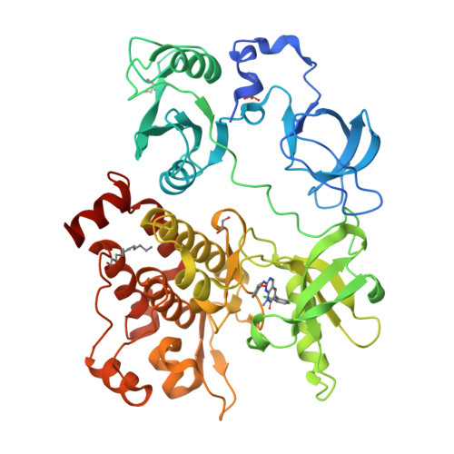
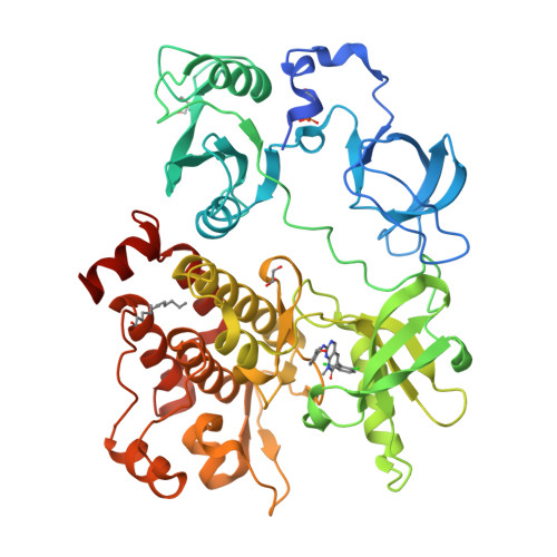
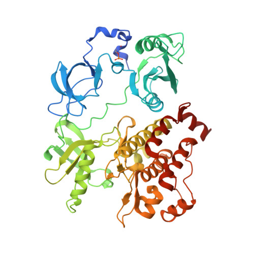
2FO0

Organization of the SH3-SH2 Unit in Active and Inactive Forms of the c-Abl Tyrosine Kinase


Experimental Data Snapshot

  • Method: X-RAY DIFFRACTION
  • Resolution: 2.27 Å
  • R-Value Free: 0.246 
  • R-Value Work: 0.210 
  • R-Value Observed: 0.210 

Starting Model: experimental
View more details

wwPDB Validation   3D Report Full Report


Ligand Structure Quality Assessment 


This is version 2.3 of the entry. See complete history


Literature

Organization of the SH3-SH2 unit in active and inactive forms of the c-Abl tyrosine kinase.

Nagar, B.Hantschel, O.Seeliger, M.Davies, J.M.Weis, W.I.Superti-Furga, G.Kuriyan, J.

(2006) Mol Cell 21: 787-798

  • DOI: https://doi.org/10.1016/j.molcel.2006.01.035
  • Primary Citation of Related Structures:  
    2FO0

  • PubMed Abstract: 

    The tyrosine kinase c-Abl is inactivated by interactions made by its SH3 and SH2 domains with the distal surface of the kinase domain. We present a crystal structure of a fragment of c-Abl which reveals that a critical N-terminal cap segment, not visualized in previous structures, buttresses the SH3-SH2 substructure in the autoinhibited state and locks it onto the distal surface of the kinase domain. Surprisingly, the N-terminal cap is phosphorylated on a serine residue that interacts with the connector between the SH3 and SH2 domains. Small-angle X-ray scattering (SAXS) analysis shows that a mutated form of c-Abl, in which the N-terminal cap and two other key contacts in the autoinhibited state are deleted, exists in an extended array of the SH3, SH2, and kinase domains. This alternative conformation of Abl is likely to prolong the active state of the kinase by preventing it from returning to the autoinhibited state.


  • Organizational Affiliation

    Howard Hughes Medical Institute, Department of Molecular and Cell Biology, University of California, Berkeley, 94720, USA.


Macromolecules
Find similar proteins by:  (by identity cutoff)  |  3D Structure
Entity ID: 1
MoleculeChains Sequence LengthOrganismDetailsImage
Proto-oncogene tyrosine-protein kinase ABL1 (1B ISOFORM)495Homo sapiensMutation(s): 2 
Gene Names: c-Abl
EC: 2.7.1.112 (PDB Primary Data), 2.7.10.2 (UniProt)
UniProt & NIH Common Fund Data Resources
Find proteins for P00519 (Homo sapiens)
Explore P00519 
Go to UniProtKB:  P00519
PHAROS:  P00519
GTEx:  ENSG00000097007 
Entity Groups  
Sequence Clusters30% Identity50% Identity70% Identity90% Identity95% Identity100% Identity
UniProt GroupP00519
Sequence Annotations
Expand
  • Reference Sequence
Small Molecules
Ligands 3 Unique
IDChains Name / Formula / InChI Key2D Diagram3D Interactions
P16
Query on P16

Download Ideal Coordinates CCD File 
C [auth A]6-(2,6-DICHLOROPHENYL)-2-{[3-(HYDROXYMETHYL)PHENYL]AMINO}-8-METHYLPYRIDO[2,3-D]PYRIMIDIN-7(8H)-ONE
C21 H16 Cl2 N4 O2
ZIQFYVPVJZEOFS-UHFFFAOYSA-N
MYR
Query on MYR

Download Ideal Coordinates CCD File 
B [auth A]MYRISTIC ACID
C14 H28 O2
TUNFSRHWOTWDNC-UHFFFAOYSA-N
GOL
Query on GOL

Download Ideal Coordinates CCD File 
D [auth A],
E [auth A]
GLYCEROL
C3 H8 O3
PEDCQBHIVMGVHV-UHFFFAOYSA-N
Modified Residues  1 Unique
IDChains TypeFormula2D DiagramParent
SEP
Query on SEP
A
L-PEPTIDE LINKINGC3 H8 N O6 PSER
Binding Affinity Annotations 
IDSourceBinding Affinity
P16 BindingDB:  2FO0 IC50: min: 2.8, max: 8 (nM) from 2 assay(s)
Experimental Data & Validation

Experimental Data

  • Method: X-RAY DIFFRACTION
  • Resolution: 2.27 Å
  • R-Value Free: 0.246 
  • R-Value Work: 0.210 
  • R-Value Observed: 0.210 
  • Space Group: P 21 21 2
Unit Cell:
Length ( Å )Angle ( ˚ )
a = 79.688α = 90
b = 117.266β = 90
c = 60.416γ = 90
Software Package:
Software NamePurpose
CNSrefinement
HKL-2000data reduction
SCALEPACKdata scaling
AMoREphasing

Structure Validation

View Full Validation Report


Ligand Structure Quality Assessment 


Entry History 

Deposition Data

Revision History  (Full details and data files)

  • Version 1.0: 2006-03-21
    Type: Initial release
  • Version 1.1: 2008-05-01
    Changes: Version format compliance
  • Version 1.2: 2011-07-13
    Changes: Non-polymer description, Version format compliance
  • Version 1.3: 2017-08-02
    Changes: Source and taxonomy
  • Version 2.0: 2021-06-30
    Changes: Advisory, Atomic model, Data collection, Database references, Derived calculations
  • Version 2.1: 2021-10-20
    Changes: Database references
  • Version 2.2: 2023-08-30
    Changes: Data collection, Refinement description
  • Version 2.3: 2024-10-30
    Changes: Structure summary