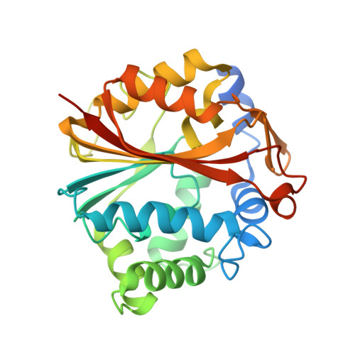
2G71

Structure of hPNMT with inhibitor 3-fluoromethyl-7-trifluoropropyl-THIQ and AdoHcy


Experimental Data Snapshot

  • Method: X-RAY DIFFRACTION
  • Resolution: 2.20 Å
  • R-Value Free: 0.251 
  • R-Value Work: 0.213 

Starting Model: experimental
View more details

wwPDB Validation   3D Report Full Report


Ligand Structure Quality Assessment 


This is version 1.5 of the entry. See complete history


Literature

Enzyme Adaptation to Inhibitor Binding: A Cryptic Binding Site in Phenylethanolamine N-Methyltransferase

Gee, C.L.Drinkwater, N.Tyndall, J.D.A.Grunewald, G.L.Wu, Q.McLeish, M.J.Martin, J.L.

(2007) J Med Chem 50: 4845-4853

  • DOI: https://doi.org/10.1021/jm0703385
  • Primary Citation of Related Structures:  
    2G70, 2G71, 2G72, 2OBF, 2ONY, 2ONZ, 2OPB

  • PubMed Abstract: 

    Shape complementarity is a fundamental principle of inhibitor design. Here we show that an enzyme for which the crystal structure has been determined (phenylethanolamine N-methyltransferase, PNMT) conceals a cryptic binding site. This site is revealed upon binding of inhibitors that are double the size of the physiological substrate. These large inhibitors are not predicted to bind in that they protrude through the accessible surface calculated from a PNMT/7-aminosulfonyl-1,2,3,4-tetrahydroisoquinoline (SK&F 29661) crystal structure, yet they are potent inhibitors of PNMT. We determined structures of the enzyme complexed with large inhibitors and found that the volume of the active site increases by 140 A3 upon binding. Changes in active site size and shape are brought about by unfavorable side chain conformations and rigid body helix motions. The energetic cost is modest, estimated at 2-3 kcal/mol from mutational analyses. Our findings further underline the importance of protein flexibility in structure-based inhibitor design studies.


  • Organizational Affiliation

    Institute for Molecular Bioscience and ARC Special Research Centre for Functional and Applied Genomics, University of Queensland, Brisbane, Qld, 4072 Australia.


Macromolecules
Find similar proteins by:  (by identity cutoff)  |  3D Structure
Entity ID: 1
MoleculeChains Sequence LengthOrganismDetailsImage
Phenylethanolamine N-methyltransferase
A, B
289Homo sapiensMutation(s): 0 
Gene Names: pnmt
EC: 2.1.1.28
UniProt & NIH Common Fund Data Resources
Find proteins for P11086 (Homo sapiens)
Explore P11086 
Go to UniProtKB:  P11086
PHAROS:  P11086
GTEx:  ENSG00000141744 
Entity Groups  
Sequence Clusters30% Identity50% Identity70% Identity90% Identity95% Identity100% Identity
UniProt GroupP11086
Sequence Annotations
Expand
  • Reference Sequence
Small Molecules
Binding Affinity Annotations 
IDSourceBinding Affinity
SAH BindingDB:  2G71 Ki: 1.40e+4 (nM) from 1 assay(s)
FTS PDBBind:  2G71 Ki: 35 (nM) from 1 assay(s)
BindingDB:  2G71 Ki: min: 35, max: 220 (nM) from 3 assay(s)
Experimental Data & Validation

Experimental Data

  • Method: X-RAY DIFFRACTION
  • Resolution: 2.20 Å
  • R-Value Free: 0.251 
  • R-Value Work: 0.213 
  • Space Group: P 43 21 2
Unit Cell:
Length ( Å )Angle ( ˚ )
a = 94.15α = 90
b = 94.15β = 90
c = 188.19γ = 90
Software Package:
Software NamePurpose
d*TREKdata processing
CNSrefinement
PDB_EXTRACTdata extraction
CrystalCleardata reduction
d*TREKdata scaling
CNSphasing

Structure Validation

View Full Validation Report


Ligand Structure Quality Assessment 


Entry History 

Deposition Data

Revision History  (Full details and data files)

  • Version 1.0: 2007-02-13
    Type: Initial release
  • Version 1.1: 2007-10-01
    Changes: Version format compliance
  • Version 1.2: 2011-07-13
    Changes: Derived calculations, Version format compliance
  • Version 1.3: 2018-04-25
    Changes: Data collection, Structure summary
  • Version 1.4: 2023-10-25
    Changes: Data collection, Database references, Derived calculations, Refinement description
  • Version 1.5: 2024-10-30
    Changes: Data collection, Structure summary