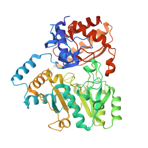
2HP1

Inter-subunit signaling in GSAM


Experimental Data Snapshot

  • Method: X-RAY DIFFRACTION
  • Resolution: 2.08 Å
  • R-Value Free: 0.254 
  • R-Value Work: 0.201 
  • R-Value Observed: 0.201 

wwPDB Validation   3D Report Full Report


Ligand Structure Quality Assessment 


This is version 1.2 of the entry. See complete history


Literature

Intersubunit signaling in glutamate-1-semialdehyde-aminomutase.

Stetefeld, J.Jenny, M.Burkhard, P.

(2006) Proc Natl Acad Sci U S A 103: 13688-13693

  • DOI: https://doi.org/10.1073/pnas.0600306103
  • Primary Citation of Related Structures:  
    2HOY, 2HOZ, 2HP1, 2HP2

  • PubMed Abstract: 

    Enzymes are highly dynamic and tightly controlled systems. However, allosteric communication linked to catalytic turnover is poorly understood. We have performed an integrated approach to trap several catalytic intermediates in the alpha2-dimeric key enzyme of chlorophyll biosynthesis, glutamate-1-semialdehyde aminomutase. Our data reveal an active-site "gating loop," which undergoes a dramatic conformational change during catalysis, that is simultaneously open in one subunit and closed in the other. This loop movement requires a beta-sheet-to-alpha-helix transition to assume the closed conformation, thus facilitating transport of substrate toward, and concomitantly forming, an integral part of the active site. The accompanying intersubunit cross-talk, which controls negative cooperativity between the allosteric pair, was explored at the atomic level. The central elements of the communication triad are the cofactor bound to different catalytic intermediates, the interface helix, and the gating loop. Together, they form a molecular switch in which the cofactor acts as a central signal transmitter linking the subunit interface with the gating loop.


  • Organizational Affiliation

    Department of Structural Biology and M. E. Müller Institute for Structural Biology, Biozentrum Universität Basel, Klingelbergstrasse 70, 4056 Basel, Switzerland. joerg.stetefeld@unibas.ch


Macromolecules
Find similar proteins by:  (by identity cutoff)  |  3D Structure
Entity ID: 1
MoleculeChains Sequence LengthOrganismDetailsImage
Glutamate-1-semialdehyde 2,1-aminomutase (GSAM) plp-form
A, B
432Synechococcus elongatus PCC 6301Mutation(s): 0 
Gene Names: hemLgsa
EC: 5.4.3.8
UniProt
Find proteins for P24630 (Synechococcus sp. (strain ATCC 27144 / PCC 6301 / SAUG 1402/1))
Explore P24630 
Go to UniProtKB:  P24630
Entity Groups  
Sequence Clusters30% Identity50% Identity70% Identity90% Identity95% Identity100% Identity
UniProt GroupP24630
Sequence Annotations
Expand
  • Reference Sequence
Small Molecules
Ligands 3 Unique
IDChains Name / Formula / InChI Key2D Diagram3D Interactions
EA5
Query on EA5

Download Ideal Coordinates CCD File 
C [auth A](4S)-4-AMINO-5-[({3-HYDROXY-2-METHYL-5-[(PHOSPHONOOXY)METHYL]PYRIDIN-4-YL}METHYL)AMINO]PENTANOIC ACID
C13 H22 N3 O7 P
DSCJVPXIMLLBAH-JTQLQIEISA-N
PLP
Query on PLP

Download Ideal Coordinates CCD File 
D [auth B]PYRIDOXAL-5'-PHOSPHATE
C8 H10 N O6 P
NGVDGCNFYWLIFO-UHFFFAOYSA-N
HOZ
Query on HOZ

Download Ideal Coordinates CCD File 
E [auth B](4S)-4,5-DIAMINOPENTANOIC ACID
C5 H12 N2 O2
PQGAAJQIFBEYSA-BYPYZUCNSA-N
Experimental Data & Validation

Experimental Data

  • Method: X-RAY DIFFRACTION
  • Resolution: 2.08 Å
  • R-Value Free: 0.254 
  • R-Value Work: 0.201 
  • R-Value Observed: 0.201 
  • Space Group: P 21 21 21
Unit Cell:
Length ( Å )Angle ( ˚ )
a = 66.483α = 90
b = 106.981β = 90
c = 119.499γ = 90
Software Package:
Software NamePurpose
CNSrefinement
MOSFLMdata reduction
CCP4data scaling
AMoREphasing

Structure Validation

View Full Validation Report


Ligand Structure Quality Assessment 


Entry History 

Deposition Data

Revision History  (Full details and data files)

  • Version 1.0: 2006-08-22
    Type: Initial release
  • Version 1.1: 2008-05-01
    Changes: Version format compliance
  • Version 1.2: 2011-07-13
    Changes: Source and taxonomy, Version format compliance