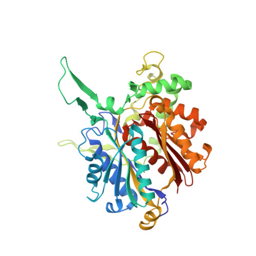
2IB7

Crystallographic and kinetic studies of human mitochondrial acetoacetyl-CoA thiolase (T2): the importance of potassium and chloride for its structure and function


Experimental Data Snapshot

  • Method: X-RAY DIFFRACTION
  • Resolution: 2.05 Å
  • R-Value Free: 0.198 
  • R-Value Work: 0.157 
  • R-Value Observed: 0.159 

Starting Model: experimental
View more details

wwPDB Validation   3D Report Full Report


This is version 1.4 of the entry. See complete history


Literature

Crystallographic and Kinetic Studies of Human Mitochondrial Acetoacetyl-CoA Thiolase: The Importance of Potassium and Chloride Ions for Its Structure and Function

Haapalainen, A.M.Merilainen, G.Pirila, P.L.Kondo, N.Fukao, T.Wierenga, R.K.

(2007) Biochemistry 46: 4305-4321

  • DOI: https://doi.org/10.1021/bi6026192
  • Primary Citation of Related Structures:  
    2IB7, 2IB8, 2IB9, 2IBU, 2IBW, 2IBY

  • PubMed Abstract: 

    Thiolases are CoA-dependent enzymes which catalyze the formation of a carbon-carbon bond in a Claisen condensation step and its reverse reaction via a thiolytic degradation mechanism. Mitochondrial acetoacetyl-coenzyme A (CoA) thiolase (T2) is important in the pathways for the synthesis and degradation of ketone bodies as well as for the degradation of 2-methylacetoacetyl-CoA. Human T2 deficiency has been identified in more than 60 patients. A unique property of T2 is its activation by potassium ions. High-resolution human T2 crystal structures are reported for the apo form and the CoA complex, with and without a bound potassium ion. The potassium ion is bound near the CoA binding site and the catalytic site. Binding of the potassium ion at this low-affinity binding site causes the rigidification of a CoA binding loop and an active site loop. Unexpectedly, a high-affinity binding site for a chloride ion has also been identified. The chloride ion is copurified, and its binding site is at the dimer interface, near two catalytic loops. A unique property of T2 is its ability to use 2-methyl-branched acetoacetyl-CoA as a substrate, whereas the other structurally characterized thiolases cannot utilize the 2-methylated compounds. The kinetic measurements show that T2 can degrade acetoacetyl-CoA and 2-methylacetoacetyl-CoA with similar catalytic efficiencies. For both substrates, the turnover numbers increase approximately 3-fold when the potassium ion concentration is increased from 0 to 40 mM KCl. The structural analysis of the active site of T2 indicates that the Phe325-Pro326 dipeptide near the catalytic cavity is responsible for the exclusive 2-methyl-branched substrate specificity.


  • Organizational Affiliation

    Biocenter Oulu and Department of Biochemistry, University of Oulu, P.O. Box 3000, FIN-90014 Oulu, Finland.


Macromolecules
Find similar proteins by:  (by identity cutoff)  |  3D Structure
Entity ID: 1
MoleculeChains Sequence LengthOrganismDetailsImage
Acetyl-CoA acetyltransferase
A, B, C, D
395Homo sapiensMutation(s): 1 
Gene Names: ACAT1
EC: 2.3.1.9
UniProt & NIH Common Fund Data Resources
Find proteins for P24752 (Homo sapiens)
Explore P24752 
Go to UniProtKB:  P24752
PHAROS:  P24752
GTEx:  ENSG00000075239 
Entity Groups  
Sequence Clusters30% Identity50% Identity70% Identity90% Identity95% Identity100% Identity
UniProt GroupP24752
Sequence Annotations
Expand
  • Reference Sequence
Small Molecules
Ligands 3 Unique
IDChains Name / Formula / InChI Key2D Diagram3D Interactions
MES
Query on MES

Download Ideal Coordinates CCD File 
F [auth A],
K [auth B],
N [auth C],
Q [auth D]
2-(N-MORPHOLINO)-ETHANESULFONIC ACID
C6 H13 N O4 S
SXGZJKUKBWWHRA-UHFFFAOYSA-N
GOL
Query on GOL

Download Ideal Coordinates CCD File 
G [auth A],
H [auth A],
I [auth A],
L [auth B],
O [auth C]
GLYCEROL
C3 H8 O3
PEDCQBHIVMGVHV-UHFFFAOYSA-N
CL
Query on CL

Download Ideal Coordinates CCD File 
E [auth A],
J [auth B],
M [auth C],
P [auth D]
CHLORIDE ION
Cl
VEXZGXHMUGYJMC-UHFFFAOYSA-M
Experimental Data & Validation

Experimental Data

  • Method: X-RAY DIFFRACTION
  • Resolution: 2.05 Å
  • R-Value Free: 0.198 
  • R-Value Work: 0.157 
  • R-Value Observed: 0.159 
  • Space Group: P 1 21 1
Unit Cell:
Length ( Å )Angle ( ˚ )
a = 75.257α = 90
b = 106.493β = 102.95
c = 101.734γ = 90
Software Package:
Software NamePurpose
REFMACrefinement
HKL-2000data collection
XDSdata reduction
XDSdata scaling
MOLREPphasing

Structure Validation

View Full Validation Report


Entry History 

Deposition Data

Revision History  (Full details and data files)

  • Version 1.0: 2007-04-03
    Type: Initial release
  • Version 1.1: 2008-05-01
    Changes: Version format compliance
  • Version 1.2: 2011-07-13
    Changes: Non-polymer description, Version format compliance
  • Version 1.3: 2021-11-10
    Changes: Data collection, Database references, Derived calculations
  • Version 1.4: 2023-10-25
    Changes: Data collection, Refinement description