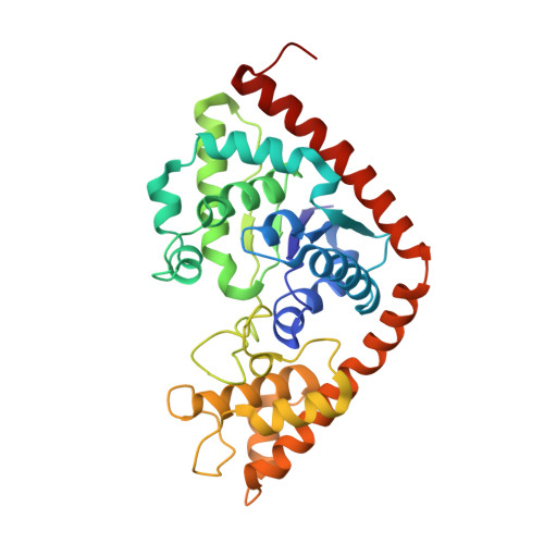
Crystal Structure of Tryptophanyl-tRNA Synthetase Complexed with Adenosine-5' Tetraphosphate: Evidence for Distributed Use of Catalytic Binding Energy in Amino Acid Activation by Class I Aminoacyl-tRNA Synthetases.
Retailleau, P., Weinreb, V., Hu, M., Carter Jr., C.W.(2007) J Mol Biol 369: 108-128
- PubMed: 17428498
- DOI: https://doi.org/10.1016/j.jmb.2007.01.091
- Primary Citation of Related Structures:
2OV4 - PubMed Abstract:
Tryptophanyl-tRNA synthetase (TrpRS) is a functionally dimeric ligase, which specifically couples hydrolysis of ATP to AMP and pyrophosphate to the formation of an ester bond between tryptophan and the cognate tRNA. TrpRS from Bacillus stearothermophilus binds the ATP analogue, adenosine-5' tetraphosphate (AQP) competitively with ATP during pyrophosphate exchange. Estimates of binding affinity from this competitive inhibition and from isothermal titration calorimetry show that AQP binds 200 times more tightly than ATP both under conditions of induced-fit, where binding is coupled to an unfavorable conformational change, and under exchange conditions, where there is no conformational change. These binding data provide an indirect experimental measurement of +3.0 kcal/mol for the conformational free energy change associated with induced-fit assembly of the active site. Thermodynamic parameters derived from the calorimetry reveal very modest enthalpic changes, consistent with binding driven largely by a favorable entropy change. The 2.5 A structure of the TrpRS:AQP complex, determined de novo by X-ray crystallography, resembles that of the previously described, pre-transition state TrpRS:ATP complexes. The anticodon-binding domain untwists relative to the Rossmann-fold domain by 20% of the way toward the orientation observed for the Products complex. An unexpected tetraphosphate conformation allows the gamma and deltad phosphate groups to occupy positions equivalent to those occupied by the beta and gamma phosphates of ATP. The beta-phosphate effects a 1.11 A extension that relocates the alpha-phosphate toward the tryptophan carboxylate while the PPi mimic moves deeper into the KMSKS loop. This configuration improves interactions between enzyme and nucleotide significantly and uniformly in the adenosine and PPi binding subsites. A new hydrogen bond forms between S194 from the class I KMSKS signature sequence and the PPi mimic. These complementary thermodynamic and structural data are all consistent with the conclusion that the tetraphosphate mimics a transition-state in which the KMSKS loop develops increasingly tight bonds to the PPi leaving group, weakening linkage to the Palpha as it is relocated by an energetically favorable domain movement. Consistent with extensive mutational data on Tyrosyl-tRNA synthetase, this aspect of the mechanism develops high transition-state affinity for the adenosine and pyrophosphate moieties, which move significantly, relative to one another, during the catalytic step.
Organizational Affiliation:
Service de Cristallochimie, ICSN-CNRS, Gif/Yvette, 91198, France.


