2OWM | pdb_00002owm

Motor domain of Neurospora crassa kinesin-3 (NcKin3)


Experimental Data Snapshot

  • Method: X-RAY DIFFRACTION
  • Resolution: 3.25 Å
  • R-Value Free: 
    0.256 (Depositor), 0.250 (DCC) 
  • R-Value Work: 
    0.210 (Depositor), 0.210 (DCC) 
  • R-Value Observed: 
    0.213 (Depositor) 

Starting Model: experimental
View more details

wwPDB Validation   3D Report Full Report


Ligand Structure Quality Assessment 

Created with Raphaël 2.3.0Worse 01 BetterLigand structure goodness of fit to experimental dataBest fitted ADPClick on this verticalbar to view details

This is version 1.3 of the entry. See complete history


Literature

X-ray Structure and Microtubule Interaction of the Motor Domain of Neurospora crassa NcKin3, a Kinesin with Unusual Processivity

Marx, A.Muller, J.Mandelkow, E.-M.Woehlke, G.Bouchet-Marquis, C.Hoenger, A.Mandelkow, E.

(2008) Biochemistry 47: 1848-1861

  • DOI: https://doi.org/10.1021/bi701483h
  • Primary Citation of Related Structures:  
    2OWM

  • PubMed Abstract: 

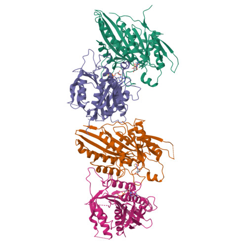
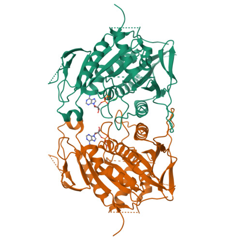
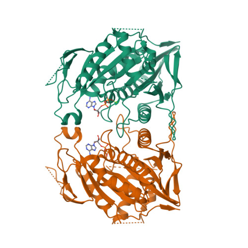
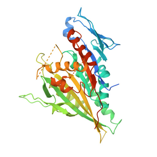
    Neurospora crassa kinesin NcKin3 belongs to a unique fungal-specific subgroup of small Kinesin-3-related motor proteins. One of its functions appears to be the transport of mitochondria along microtubules. Here, we present the X-ray structure of a C-terminally truncated monomeric construct of NcKin3 comprising the motor domain and the neck linker, and a 3-D image reconstruction of this motor domain bound to microtubules, by cryoelectron microscopy. The protein contains Mg.ADP bound to the active site, yet the structure resembles an ATP-bound state. By comparison with structures of the Kinesin-3 motor Kif1A in different nucleotide states (Kikkawa, M. et al. (2001) Nature (London, U.K.) 411, 439-445), the NcKin3 structure corresponds to the AMPPCP complex of Kif1A rather than the AMPPNP complex. NcKin3-specific differences in the coordination of the nucleotide and asymmetric interactions between adjacent molecules in the crystal are discussed in the context of the unusual kinetics of the dimeric wild-type motor and the monomeric construct used for crystal structure analysis. The NcKin3 motor decorates microtubules at a stoichiometry of one head per alphabeta-tubulin heterodimer, thereby forming an axial periodicity of 8 nm. In spite of unusual extensions at the N-terminus and within flexible loops L2, L8a, and L12 (corresponding to the K-loop of monomeric kinesins), the microtubule binding geometry is similar to that of other members of the kinesin family.


  • Organizational Affiliation

    Max-Planck-Unit for Structural Molecular Biology, Notkestrasse 85, 22607 Hamburg, Germany. marx@mpasmb.desy.de


Macromolecules
Find similar proteins by:  (by identity cutoff)  |  3D Structure
Entity ID: 1
MoleculeChains Sequence LengthOrganismDetailsImage
Related to KINESIN-LIKE PROTEIN KIF1C
A, B, C, D
443Neurospora crassaMutation(s): 0 
Gene Names: 17E5.250
EC: 3.6.4.4
UniProt
Find proteins for Q9C2M3 (Neurospora crassa)
Explore Q9C2M3 
Go to UniProtKB:  Q9C2M3
Entity Groups  
Sequence Clusters30% Identity50% Identity70% Identity90% Identity95% Identity100% Identity
UniProt GroupQ9C2M3
Sequence Annotations
Expand
  • Reference Sequence
Small Molecules
Ligands 2 Unique
IDChains Name / Formula / InChI Key2D Diagram3D Interactions
ADP
Query on ADP

Download Ideal Coordinates CCD File 
F [auth A],
H [auth B],
J [auth C],
L [auth D]
ADENOSINE-5'-DIPHOSPHATE
C10 H15 N5 O10 P2
XTWYTFMLZFPYCI-KQYNXXCUSA-N
MG
Query on MG

Download Ideal Coordinates CCD File 
E [auth A],
G [auth B],
I [auth C],
K [auth D]
MAGNESIUM ION
Mg
JLVVSXFLKOJNIY-UHFFFAOYSA-N
Experimental Data & Validation

Experimental Data

  • Method: X-RAY DIFFRACTION
  • Resolution: 3.25 Å
  • R-Value Free:  0.256 (Depositor), 0.250 (DCC) 
  • R-Value Work:  0.210 (Depositor), 0.210 (DCC) 
  • R-Value Observed: 0.213 (Depositor) 
Space Group: P 1 21 1
Unit Cell:
Length ( Å )Angle ( ˚ )
a = 75.945α = 90
b = 98.408β = 91.86
c = 111.764γ = 90
Software Package:
Software NamePurpose
DENZOdata reduction
SCALEPACKdata scaling
PHASERphasing
REFMACrefinement
PDB_EXTRACTdata extraction

Structure Validation

View Full Validation Report


Ligand Structure Quality Assessment 

Created with Raphaël 2.3.0Worse 01 BetterLigand structure goodness of fit to experimental dataBest fitted ADPClick on this verticalbar to view details

Entry History 

Deposition Data

Revision History  (Full details and data files)

  • Version 1.0: 2008-01-29
    Type: Initial release
  • Version 1.1: 2011-07-13
    Changes: Advisory, Refinement description, Version format compliance
  • Version 1.2: 2017-10-18
    Changes: Refinement description
  • Version 1.3: 2023-08-30
    Changes: Data collection, Database references, Derived calculations, Refinement description