2QKM

The crystal structure of fission yeast mRNA decapping enzyme Dcp1-Dcp2 complex


Experimental Data Snapshot

  • Method: X-RAY DIFFRACTION
  • Resolution: 2.80 Å
  • R-Value Free: 0.289 
  • R-Value Work: 0.233 
  • R-Value Observed: 0.236 

Starting Models: experimental
View more details

wwPDB Validation   3D Report Full Report


Ligand Structure Quality Assessment 


This is version 1.2 of the entry. See complete history


Literature

Structural basis of dcp2 recognition and activation by dcp1.

She, M.Decker, C.J.Svergun, D.I.Round, A.Chen, N.Muhlrad, D.Parker, R.Song, H.

(2008) Mol Cell 29: 337-349

  • DOI: https://doi.org/10.1016/j.molcel.2008.01.002
  • Primary Citation of Related Structures:  
    2QKL, 2QKM

  • PubMed Abstract: 

    A critical step in mRNA degradation is the removal of the 5' cap structure, which is catalyzed by the Dcp1-Dcp2 complex. The crystal structure of an S. pombe Dcp1p-Dcp2n complex combined with small-angle X-ray scattering analysis (SAXS) reveals that Dcp2p exists in open and closed conformations, with the closed complex being, or closely resembling, the catalytically more active form. This suggests that a conformational change between these open and closed complexes might control decapping. A bipartite RNA-binding channel containing the catalytic site and Box B motif is identified with a bound ATP located in the catalytic pocket in the closed complex, suggesting possible interactions that facilitate substrate binding. Dcp1 stimulates the activity of Dcp2 by promoting and/or stabilizing the closed complex. Notably, the interface of Dcp1 and Dcp2 is not fully conserved, explaining why the Dcp1-Dcp2 interaction in higher eukaryotes requires an additional factor.


  • Organizational Affiliation

    Laboratory of Macromolecular Structure, Institute of Molecular and Cell Biology, Agency for Science, Technology and Research, 61 Biopolis Drive, Proteos, 138673 Singapore.


Macromolecules
Find similar proteins by:  (by identity cutoff)  |  3D Structure
Entity ID: 1
MoleculeChains Sequence LengthOrganismDetailsImage
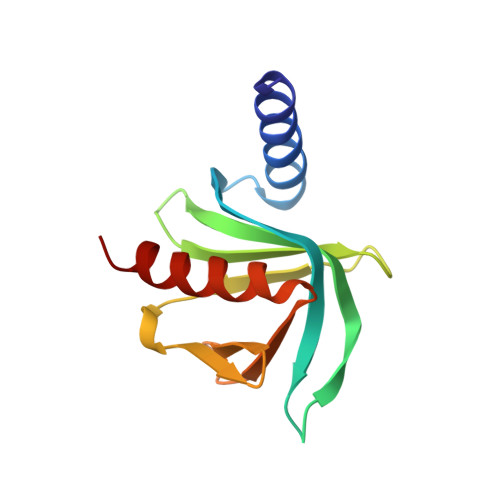
SPBC3B9.21 protein
A, C, E, G
127Schizosaccharomyces pombeMutation(s): 0 
Gene Names: dcp1
EC: 3.6.1.30
UniProt
Find proteins for Q9P805 (Schizosaccharomyces pombe (strain 972 / ATCC 24843))
Explore Q9P805 
Go to UniProtKB:  Q9P805
Entity Groups  
Sequence Clusters30% Identity50% Identity70% Identity90% Identity95% Identity100% Identity
UniProt GroupQ9P805
Sequence Annotations
Expand
  • Reference Sequence
Find similar proteins by:  (by identity cutoff)  |  3D Structure
Entity ID: 2
MoleculeChains Sequence LengthOrganismDetailsImage
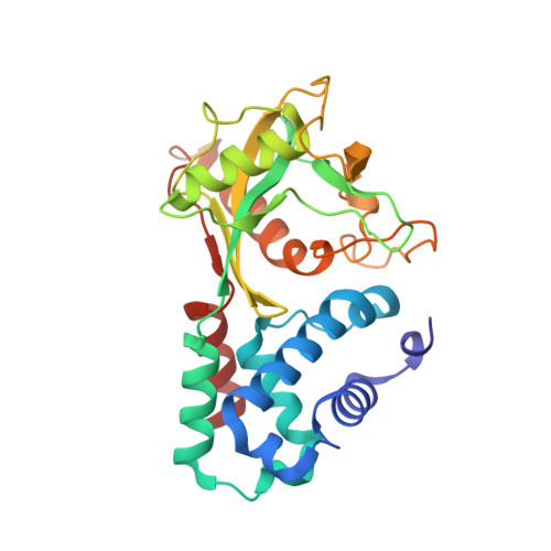
SPAC19A8.12 protein
B, D, F, H
266Schizosaccharomyces pombeMutation(s): 0 
Gene Names: SPAC19A8.12
EC: 3
UniProt
Find proteins for O13828 (Schizosaccharomyces pombe (strain 972 / ATCC 24843))
Explore O13828 
Go to UniProtKB:  O13828
Entity Groups  
Sequence Clusters30% Identity50% Identity70% Identity90% Identity95% Identity100% Identity
UniProt GroupO13828
Sequence Annotations
Expand
  • Reference Sequence
Experimental Data & Validation

Experimental Data

  • Method: X-RAY DIFFRACTION
  • Resolution: 2.80 Å
  • R-Value Free: 0.289 
  • R-Value Work: 0.233 
  • R-Value Observed: 0.236 
  • Space Group: P 1 21 1
Unit Cell:
Length ( Å )Angle ( ˚ )
a = 68.289α = 90
b = 161.443β = 97.49
c = 91.386γ = 90
Software Package:
Software NamePurpose
REFMACrefinement
PDB_EXTRACTdata extraction
ADSCdata collection
MOSFLMdata reduction
SCALAdata scaling
PHASESphasing

Structure Validation

View Full Validation Report


Ligand Structure Quality Assessment 


Entry History 

Deposition Data

Revision History  (Full details and data files)

  • Version 1.0: 2008-04-15
    Type: Initial release
  • Version 1.1: 2011-07-13
    Changes: Advisory, Version format compliance
  • Version 1.2: 2023-08-30
    Changes: Data collection, Database references, Derived calculations, Refinement description