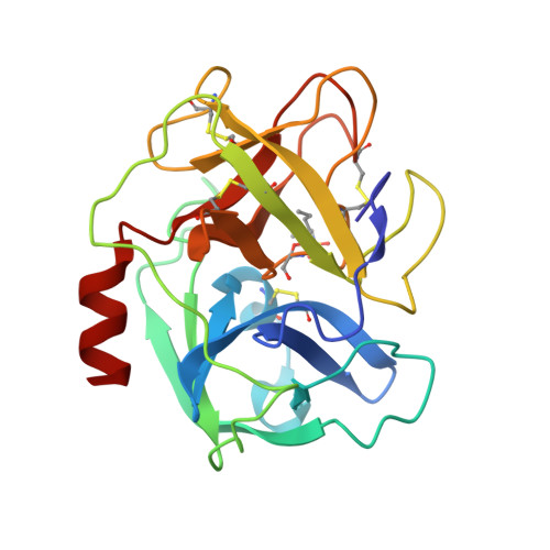
2RG3

Covalent complex structure of elastase


Experimental Data Snapshot

  • Method: X-RAY DIFFRACTION
  • Resolution: 1.80 Å
  • R-Value Work: 0.200 

wwPDB Validation   3D Report Full Report


This is version 2.0 of the entry. See complete history


Literature

X-ray snapshot of the mechanism of inactivation of human neutrophil elastase by 1,2,5-thiadiazolidin-3-one 1,1-dioxide derivatives.

Huang, W.Yamamoto, Y.Li, Y.Dou, D.Alliston, K.R.Hanzlik, R.P.Williams, T.D.Groutas, W.C.

(2008) J Med Chem 51: 2003-2008

  • DOI: https://doi.org/10.1021/jm700966p
  • Primary Citation of Related Structures:  
    2RG3

  • PubMed Abstract: 

    The mechanism of action of a general class of mechanism-based inhibitors of serine proteases, including human neutrophil elastase (HNE), has been elucidated by determining the X-ray crystal structure of an enzyme-inhibitor complex. The captured intermediate indicates that processing of inhibitor by the enzyme generates an N-sulfonyl imine functionality that is tethered to Ser195, in accordance with the postulated mechanism of action of this class of inhibitors. The identity of the HNE-N-sulfonyl imine species was further corroborated using electrospray ionization mass spectrometry.


  • Organizational Affiliation

    Department of Chemistry, Wichita State University, Wichita, KS 67260, USA.


Macromolecules
Find similar proteins by:  (by identity cutoff)  |  3D Structure
Entity ID: 1
MoleculeChains Sequence LengthOrganismDetailsImage
Leukocyte elastase218Homo sapiensMutation(s): 1 
Gene Names: ELA2
EC: 3.4.21.37
UniProt & NIH Common Fund Data Resources
Find proteins for P08246 (Homo sapiens)
Explore P08246 
Go to UniProtKB:  P08246
PHAROS:  P08246
GTEx:  ENSG00000197561 
Entity Groups  
Sequence Clusters30% Identity50% Identity70% Identity90% Identity95% Identity100% Identity
UniProt GroupP08246
Glycosylation
Glycosylation Sites: 1Go to GlyGen: P08246-1
Sequence Annotations
Expand
  • Reference Sequence
Oligosaccharides

Help

Entity ID: 2
MoleculeChains Length2D Diagram Glycosylation3D Interactions
2-acetamido-2-deoxy-beta-D-glucopyranose-(1-4)-[alpha-L-fucopyranose-(1-6)]2-acetamido-2-deoxy-beta-D-glucopyranose
B
3N-Glycosylation
Glycosylation Resources
GlyTouCan:  G21290RB
GlyCosmos:  G21290RB
GlyGen:  G21290RB
Small Molecules
Ligands 1 Unique
IDChains Name / Formula / InChI Key2D Diagram3D Interactions
EPE
Query on EPE

Download Ideal Coordinates CCD File 
C [auth A]4-(2-HYDROXYETHYL)-1-PIPERAZINE ETHANESULFONIC ACID
C8 H18 N2 O4 S
JKMHFZQWWAIEOD-UHFFFAOYSA-N
Modified Residues  1 Unique
IDChains TypeFormula2D DiagramParent
BG1
Query on BG1
A
L-PEPTIDE LINKINGC10 H21 N3 O6 SSER
Experimental Data & Validation

Experimental Data

  • Method: X-RAY DIFFRACTION
  • Resolution: 1.80 Å
  • R-Value Work: 0.200 
  • Space Group: P 63
Unit Cell:
Length ( Å )Angle ( ˚ )
a = 73.2α = 90
b = 73.2β = 90
c = 70.1γ = 120
Software Package:
Software NamePurpose
HKL-2000data collection
PHASERphasing
CNSrefinement
HKL-2000data reduction
HKL-2000data scaling

Structure Validation

View Full Validation Report


Entry History 

Deposition Data

Revision History  (Full details and data files)

  • Version 1.0: 2008-07-01
    Type: Initial release
  • Version 1.1: 2011-07-13
    Changes: Non-polymer description, Version format compliance
  • Version 2.0: 2020-07-29
    Type: Remediation
    Reason: Carbohydrate remediation
    Changes: Atomic model, Data collection, Database references, Derived calculations, Structure summary