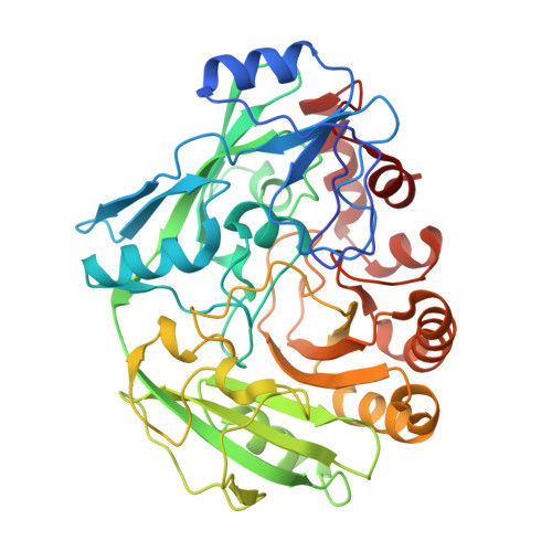
2VFT

Alditol Oxidase from Streptomyces coelicolor A3(2): Complex with Sorbitol


Experimental Data Snapshot

  • Method: X-RAY DIFFRACTION
  • Resolution: 1.60 Å
  • R-Value Free: 0.179 
  • R-Value Work: 0.152 
  • R-Value Observed: 0.154 

Starting Model: experimental
View more details

wwPDB Validation   3D Report Full Report


Ligand Structure Quality Assessment 


This is version 1.4 of the entry. See complete history


Literature

Structural Analysis of the Catalytic Mechanism and Stereoselectivity in Streptomyces Coelicolor Alditol Oxidase.

Forneris, F.Heuts, D.P.H.M.Delvecchio, M.Rovida, S.Fraaije, M.W.Mattevi, A.

(2008) Biochemistry 47: 978

  • DOI: https://doi.org/10.1021/bi701886t
  • Primary Citation of Related Structures:  
    2VFR, 2VFS, 2VFT, 2VFU, 2VFV

  • PubMed Abstract: 

    Alditol oxidase (AldO) from Streptomyces coelicolor A3(2) is a soluble monomeric flavin-dependent oxidase that performs selective oxidation of the terminal primary hydroxyl group of several alditols. Here, we report the crystal structure of the recombinant enzyme in its native state and in complex with both six-carbon (mannitol and sorbitol) and five-carbon substrates (xylitol). AldO shares the same folding topology of the members of the vanillyl-alcohol oxidase family of flavoenzymes and exhibits a covalently linked FAD which is located at the bottom of a funnel-shaped pocket that forms the active site. The high resolution of the three-dimensional structures highlights a well-defined hydrogen-bonding network that tightly constrains the substrate in the productive conformation for catalysis. Substrate binding occurs through a lock-and-key mechanism and does not induce conformational changes with respect to the ligand-free protein. A network of charged residues is proposed to favor catalysis through stabilization of the deprotonated form of the substrate. A His side chain acts as back door that "pushes" the substrate-reactive carbon atom toward the N5-C4a locus of the flavin. Analysis of the three-dimensional structure reveals possible pathways for diffusion of molecular oxygen and a small cavity on the re side of the flavin that may host oxygen during FAD reoxidation. These features combined with the tight shape of the catalytic site provide insights into the mechanism of AldO-mediated regioselective oxidation reactions and its substrate specificity.


  • Organizational Affiliation

    Department of Genetics and Microbiology, University of Pavia, Via Ferrata 1, 27100 Pavia, Italy.


Macromolecules
Find similar proteins by:  (by identity cutoff)  |  3D Structure
Entity ID: 1
MoleculeChains Sequence LengthOrganismDetailsImage
XYLITOL OXIDASE422Streptomyces coelicolor A3(2)Mutation(s): 0 
EC: 1.1.3.41
UniProt
Find proteins for Q9ZBU1 (Streptomyces coelicolor (strain ATCC BAA-471 / A3(2) / M145))
Explore Q9ZBU1 
Go to UniProtKB:  Q9ZBU1
Entity Groups  
Sequence Clusters30% Identity50% Identity70% Identity90% Identity95% Identity100% Identity
UniProt GroupQ9ZBU1
Sequence Annotations
Expand
  • Reference Sequence
Small Molecules
Ligands 3 Unique
IDChains Name / Formula / InChI Key2D Diagram3D Interactions
FAD
Query on FAD

Download Ideal Coordinates CCD File 
B [auth A]FLAVIN-ADENINE DINUCLEOTIDE
C27 H33 N9 O15 P2
VWWQXMAJTJZDQX-UYBVJOGSSA-N
SOR
Query on SOR

Download Ideal Coordinates CCD File 
C [auth A]sorbitol
C6 H14 O6
FBPFZTCFMRRESA-JGWLITMVSA-N
CL
Query on CL

Download Ideal Coordinates CCD File 
D [auth A]CHLORIDE ION
Cl
VEXZGXHMUGYJMC-UHFFFAOYSA-M
Experimental Data & Validation

Experimental Data

  • Method: X-RAY DIFFRACTION
  • Resolution: 1.60 Å
  • R-Value Free: 0.179 
  • R-Value Work: 0.152 
  • R-Value Observed: 0.154 
  • Space Group: C 1 2 1
Unit Cell:
Length ( Å )Angle ( ˚ )
a = 107.185α = 90
b = 66.622β = 95.09
c = 59.134γ = 90
Software Package:
Software NamePurpose
REFMACrefinement
MOSFLMdata reduction
CCP4data scaling
CCP4phasing

Structure Validation

View Full Validation Report


Ligand Structure Quality Assessment 


Entry History 

Deposition Data

Revision History  (Full details and data files)

  • Version 1.0: 2008-01-08
    Type: Initial release
  • Version 1.1: 2011-05-08
    Changes: Version format compliance
  • Version 1.2: 2011-07-13
    Changes: Version format compliance
  • Version 1.3: 2020-07-29
    Type: Remediation
    Reason: Carbohydrate remediation
    Changes: Derived calculations, Other, Structure summary
  • Version 1.4: 2023-12-13
    Changes: Data collection, Database references, Refinement description, Structure summary