2VHF | pdb_00002vhf

Structure of the CYLD USP domain


Experimental Data Snapshot

  • Method: X-RAY DIFFRACTION
  • Resolution: 2.80 Å
  • R-Value Free: 
    0.281 (Depositor), 0.280 (DCC) 
  • R-Value Work: 
    0.233 (Depositor), 0.240 (DCC) 
  • R-Value Observed: 
    0.235 (Depositor) 

wwPDB Validation   3D Report Full Report


This is version 1.3 of the entry. See complete history


Literature

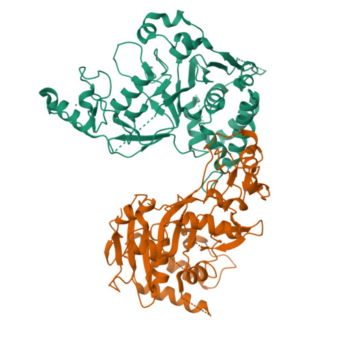
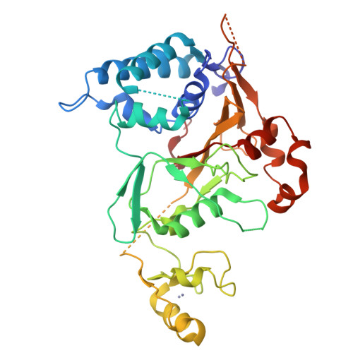
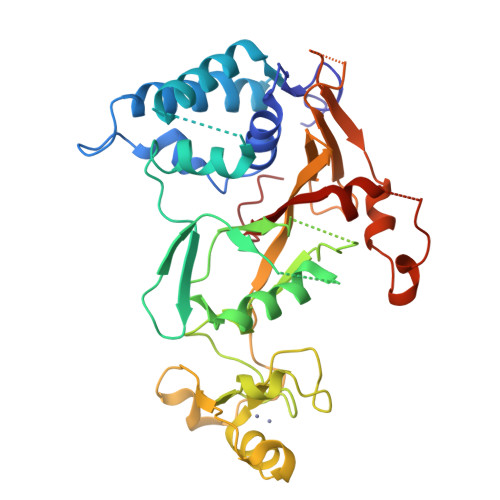
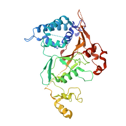
The Structure of the Cyld Usp Domain Explains its Specificity for Lys63-Linked Polyubiquitin and Reveals a B-Box Module

Komander, D.Lord, C.J.Scheel, H.Swift, S.Hofmann, K.Ashworth, A.Barford, D.

(2008) Mol Cell Biol 29: 451

  • DOI: https://doi.org/10.1016/j.molcel.2007.12.018
  • Primary Citation of Related Structures:  
    2VHF

  • PubMed Abstract: 

    The tumor suppressor CYLD antagonizes NF-kappaB and JNK signaling by disassembly of Lys63-linked ubiquitin chains synthesized in response to cytokine stimulation. Here we describe the crystal structure of the CYLD USP domain, revealing a distinctive architecture that provides molecular insights into its specificity toward Lys63-linked polyubiquitin. We identify regions of the USP domain responsible for this specificity and demonstrate endodeubiquitinase activity toward such chains. Pathogenic truncations of the CYLD C terminus, associated with the hypertrophic skin tumor cylindromatosis, disrupt the USP domain, accounting for loss of CYLD catalytic activity. A small zinc-binding B box domain, similar in structure to other crossbrace Zn-binding folds--including the RING domain found in E3 ubiquitin ligases--is inserted within the globular core of the USP domain. Biochemical and functional characterization of the B box suggests a role as a protein-interaction module that contributes to determining the subcellular localization of CYLD.


  • Organizational Affiliation

    Section of Structural Biology, Institute of Cancer Research, Chester Beatty Laboratories, 237 Fulham Road, London SW3 6JB, UK.


Macromolecules
Find similar proteins by:  (by identity cutoff)  |  3D Structure
Entity ID: 1
MoleculeChains Sequence LengthOrganismDetailsImage
UBIQUITIN CARBOXYL-TERMINAL HYDROLASE CYLD
A, B
374Homo sapiensMutation(s): 0 
EC: 3.1.2.15 (PDB Primary Data), 3.4.19.12 (UniProt)
UniProt & NIH Common Fund Data Resources
Find proteins for Q9NQC7 (Homo sapiens)
Explore Q9NQC7 
Go to UniProtKB:  Q9NQC7
PHAROS:  Q9NQC7
GTEx:  ENSG00000083799 
Entity Groups  
Sequence Clusters30% Identity50% Identity70% Identity90% Identity95% Identity100% Identity
UniProt GroupQ9NQC7
Sequence Annotations
Expand
  • Reference Sequence
Experimental Data & Validation

Experimental Data

  • Method: X-RAY DIFFRACTION
  • Resolution: 2.80 Å
  • R-Value Free:  0.281 (Depositor), 0.280 (DCC) 
  • R-Value Work:  0.233 (Depositor), 0.240 (DCC) 
  • R-Value Observed: 0.235 (Depositor) 
Space Group: P 21 21 21
Unit Cell:
Length ( Å )Angle ( ˚ )
a = 60.494α = 90
b = 89.083β = 90
c = 171.806γ = 90
Software Package:
Software NamePurpose
REFMACrefinement
MOSFLMdata reduction
SCALAdata scaling
SHELXCDEphasing
HKL2Mapphasing
SHARPphasing

Structure Validation

View Full Validation Report


Entry History 

Deposition Data

Revision History  (Full details and data files)

  • Version 1.0: 2008-03-11
    Type: Initial release
  • Version 1.1: 2011-05-08
    Changes: Version format compliance
  • Version 1.2: 2011-07-13
    Changes: Version format compliance
  • Version 1.3: 2024-05-08
    Changes: Data collection, Database references, Derived calculations, Other, Refinement description