2W8H | pdb_00002w8h

Crystal structure of spin labeled Wza24-345.


Experimental Data Snapshot

  • Method: X-RAY DIFFRACTION
  • Resolution: 2.76 Å
  • R-Value Free: 
    0.229 (Depositor), 0.220 (DCC) 
  • R-Value Work: 
    0.211 (Depositor), 0.210 (DCC) 
  • R-Value Observed: 
    0.212 (Depositor) 

Starting Model: experimental
View more details

wwPDB Validation 


Ligand Structure Quality Assessment 

Created with Raphaël 2.3.0Worse 01 BetterLigand structure goodness of fit to experimental dataBest fitted MTNClick on this verticalbar to view details

This is version 1.4 of the entry. See complete history


Literature

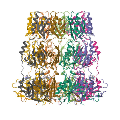
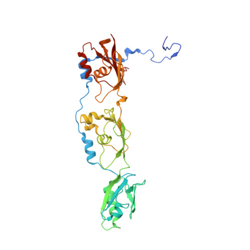
Peldor Distance Fingerprinting of the Octameric Outer-Membrane Protein Wza from Escherichia Coli.

Hagelueken, G.Ingledew, W.J.Huang, H.Petrovic-Stojanovska, B.Whitfield, C.Elmkami, H.Schiemann, O.Naismith, J.H.

(2009) Angew Chem Int Ed Engl 48: 2904

  • DOI: https://doi.org/10.1002/anie.200805758
  • Primary Citation of Related Structures:  
    2W8H, 2W8I

  • PubMed Abstract: 

    Distance fingerprinting: Pulsed electron-electron double resonance spectroscopy (PELDOR) is applied to the octameric membrane protein complex Wza of E. coli. The data yielded a detailed distance fingerprint of its periplasmic region that compares favorably to the crystal structure. These results provide the foundation to study conformation changes from interaction with partner proteins.


  • Organizational Affiliation

    Centre for Biomolecular Sciences, The University of St. Andrews, Fife KY16 9RH, UK.


Macromolecules
Find similar proteins by:  (by identity cutoff)  |  3D Structure
Entity ID: 1
MoleculeChains Sequence LengthOrganismDetailsImage
PUTATIVE OUTER MEMBRANE LIPOPROTEIN WZA
A, B, C, D, E
329Escherichia coliMutation(s): 1 
Membrane Entity: Yes 
UniProt
Find proteins for Q9X4B7 (Escherichia coli)
Explore Q9X4B7 
Go to UniProtKB:  Q9X4B7
Entity Groups  
Sequence Clusters30% Identity50% Identity70% Identity90% Identity95% Identity100% Identity
UniProt GroupQ9X4B7
Sequence Annotations
Expand
  • Reference Sequence
Small Molecules
Ligands 2 Unique
IDChains Name / Formula / InChI Key2D Diagram3D Interactions
MTN
Query on MTN

Download Ideal Coordinates CCD File 
I [auth A]
J [auth B]
K [auth C]
L [auth D]
N [auth E]
S-[(1-oxyl-2,2,5,5-tetramethyl-2,5-dihydro-1H-pyrrol-3-yl)methyl] methanesulfonothioate
C10 H18 N O3 S2
MXZPGYFBZHBAQM-UHFFFAOYSA-N
CL
Query on CL

Download Ideal Coordinates CCD File 
M [auth D]CHLORIDE ION
Cl
VEXZGXHMUGYJMC-UHFFFAOYSA-M
Experimental Data & Validation

Experimental Data

  • Method: X-RAY DIFFRACTION
  • Resolution: 2.76 Å
  • R-Value Free:  0.229 (Depositor), 0.220 (DCC) 
  • R-Value Work:  0.211 (Depositor), 0.210 (DCC) 
  • R-Value Observed: 0.212 (Depositor) 
Space Group: P 21 21 2
Unit Cell:
Length ( Å )Angle ( ˚ )
a = 141.153α = 90
b = 141.152β = 90
c = 167.529γ = 90
Software Package:
Software NamePurpose
PHENIXrefinement
HKL-2000data reduction
HKL-2000data scaling
PHASERphasing

Structure Validation


Ligand Structure Quality Assessment 

Created with Raphaël 2.3.0Worse 01 BetterLigand structure goodness of fit to experimental dataBest fitted MTNClick on this verticalbar to view details

Entry History 

Deposition Data

Revision History  (Full details and data files)

  • Version 1.0: 2009-02-10
    Type: Initial release
  • Version 1.1: 2011-05-12
    Changes: Version format compliance
  • Version 1.2: 2011-07-13
    Changes: Version format compliance
  • Version 1.3: 2023-12-13
    Changes: Advisory, Data collection, Database references, Derived calculations, Other, Refinement description
  • Version 1.4: 2024-10-16
    Changes: Structure summary