Serendipitous Fatty Acid Binding Reveals the Structural Determinants for Ligand Recognition in Apolipoprotein M.
Sevvana, M., Ahnstrom, J., Egerer-Sieber, C., Lange, H.A., Dahlback, B., Muller, Y.A.(2009) J Mol Biol 393: 920
- PubMed: 19733574
- DOI: https://doi.org/10.1016/j.jmb.2009.08.071
- Primary Citation of Related Structures:
2WEW, 2WEX - PubMed Abstract:
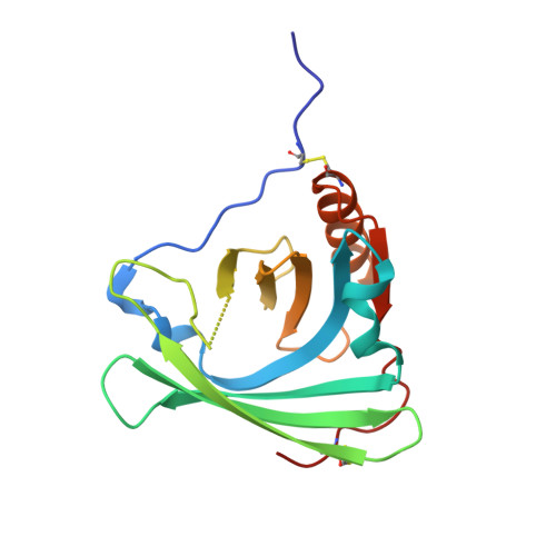
Apolipoprotein M (ApoM) is a 25-kDa HDL-associated apolipoprotein and a member of the lipocalin family of proteins. Mature apoM retains its signal peptide, which serves as a lipid anchor attaching apoM to the lipoproteins, thereby keeping it in the circulation. Studies in mice have suggested apoM to be antiatherogenic, but its physiological function is yet unknown. We have now determined the 1.95 A resolution crystal structure of recombinant human apoM expressed in Escherichia coli and made the unexpected discovery that apoM, although refolded from inclusion bodies, was in complex with fatty acids containing 14, 16 or 18 carbon atoms. ApoM displays the typical lipocalin fold characterised by an eight-stranded antiparallel beta-barrel that encloses an internal ligand-binding pocket. The crystal structures of two different complexes provide a detailed picture of the ligand-binding determinants of apoM. Additional fatty acid- and lipid-binding studies with apoM and the mutants apoM(W47F) and apoM(W100F) showed that sphingosine-1-phosphate is able to displace the bound fatty acids and efficiently quenched the intrinsic fluorescence with an IC(50) of 0.90 muM. Whereas the fatty acids bound in the crystal structure could be a mere consequence of recombinant protein production, the observed binding of sphingosine-1-phosphate might provide a key to a better understanding of the physiological function of apoM.
Organizational Affiliation:
Lehrstuhl für Biotechnik, Department of Biology, Friedrich-Alexander-University Erlangen-Nuremberg, Im IZMP, Henkestr. 91, D-91052 Erlangen, Germany.


