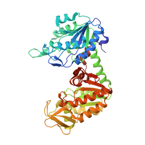
2XE7

The complete reaction cycle of human phosphoglycerate kinase: The open ternary complex with 3PG and ADP


Experimental Data Snapshot

  • Method: X-RAY DIFFRACTION
  • Resolution: 2.20 Å
  • R-Value Free: 0.288 
  • R-Value Work: 0.224 
  • R-Value Observed: 0.228 

Starting Model: experimental
View more details

wwPDB Validation   3D Report Full Report


Ligand Structure Quality Assessment 


This is version 1.2 of the entry. See complete history


Literature

A Spring Loaded Release Mechanism Regulates Domain Movement and Catalysis in Phosphoglycerate Kinase.

Zerrad, L.Merli, A.Schroder, G.F.Varga, A.Graczer, E.Pernot, P.Round, A.Vas, M.Bowler, M.W.

(2011) J Biol Chem 286: 14040

  • DOI: https://doi.org/10.1074/jbc.M110.206813
  • Primary Citation of Related Structures:  
    2XE6, 2XE7, 2XE8

  • PubMed Abstract: 

    Phosphoglycerate kinase (PGK) is the enzyme responsible for the first ATP-generating step of glycolysis and has been implicated extensively in oncogenesis and its development. Solution small angle x-ray scattering (SAXS) data, in combination with crystal structures of the enzyme in complex with substrate and product analogues, reveal a new conformation for the resting state of the enzyme and demonstrate the role of substrate binding in the preparation of the enzyme for domain closure. Comparison of the x-ray scattering curves of the enzyme in different states with crystal structures has allowed the complete reaction cycle to be resolved both structurally and temporally. The enzyme appears to spend most of its time in a fully open conformation with short periods of closure and catalysis, thereby allowing the rapid diffusion of substrates and products in and out of the binding sites. Analysis of the open apoenzyme structure, defined through deformable elastic network refinement against the SAXS data, suggests that interactions in a mostly buried hydrophobic region may favor the open conformation. This patch is exposed on domain closure, making the open conformation more thermodynamically stable. Ionic interactions act to maintain the closed conformation to allow catalysis. The short time PGK spends in the closed conformation and its strong tendency to rest in an open conformation imply a spring-loaded release mechanism to regulate domain movement, catalysis, and efficient product release.


  • Organizational Affiliation

    Structural Biology Group, European Synchrotron Radiation Facility, 6 rue Jules Horowitz, F-38043 Grenoble, France.


Macromolecules
Find similar proteins by:  (by identity cutoff)  |  3D Structure
Entity ID: 1
MoleculeChains Sequence LengthOrganismDetailsImage
PHOSPHOGLYCERATE KINASE 1417Homo sapiensMutation(s): 0 
EC: 2.7.2.3
UniProt & NIH Common Fund Data Resources
Find proteins for P00558 (Homo sapiens)
Explore P00558 
Go to UniProtKB:  P00558
PHAROS:  P00558
GTEx:  ENSG00000102144 
Entity Groups  
Sequence Clusters30% Identity50% Identity70% Identity90% Identity95% Identity100% Identity
UniProt GroupP00558
Sequence Annotations
Expand
  • Reference Sequence
Small Molecules
Ligands 2 Unique
IDChains Name / Formula / InChI Key2D Diagram3D Interactions
ADP
Query on ADP

Download Ideal Coordinates CCD File 
C [auth A]ADENOSINE-5'-DIPHOSPHATE
C10 H15 N5 O10 P2
XTWYTFMLZFPYCI-KQYNXXCUSA-N
3PG
Query on 3PG

Download Ideal Coordinates CCD File 
B [auth A]3-PHOSPHOGLYCERIC ACID
C3 H7 O7 P
OSJPPGNTCRNQQC-UWTATZPHSA-N
Binding Affinity Annotations 
IDSourceBinding Affinity
3PG BindingDB:  2XE7 Kd: 1.09e+5 (nM) from 1 assay(s)
Experimental Data & Validation

Experimental Data

  • Method: X-RAY DIFFRACTION
  • Resolution: 2.20 Å
  • R-Value Free: 0.288 
  • R-Value Work: 0.224 
  • R-Value Observed: 0.228 
  • Space Group: P 21 21 2
Unit Cell:
Length ( Å )Angle ( ˚ )
a = 58.62α = 90
b = 73.45β = 90
c = 93.44γ = 90
Software Package:
Software NamePurpose
REFMACrefinement
MOSFLMdata reduction
SCALAdata scaling
MOLREPphasing

Structure Validation

View Full Validation Report


Ligand Structure Quality Assessment 


Entry History 

Deposition Data

Revision History  (Full details and data files)

  • Version 1.0: 2011-01-19
    Type: Initial release
  • Version 1.1: 2011-10-26
    Changes: Database references, Version format compliance
  • Version 1.2: 2023-12-20
    Changes: Data collection, Database references, Derived calculations, Other, Refinement description