2XT3 | pdb_00002xt3

HUMAN KIF7, A KINESIN INVOLVED IN HEDGEHOG SIGNALLING


Experimental Data Snapshot

  • Method: X-RAY DIFFRACTION
  • Resolution: 1.88 Å
  • R-Value Free: 
    0.229 (Depositor), 0.230 (DCC) 
  • R-Value Work: 
    0.179 (Depositor), 0.180 (DCC) 
  • R-Value Observed: 
    0.182 (Depositor) 

Starting Model: experimental
View more details

wwPDB Validation   3D Report Full Report


Ligand Structure Quality Assessment 

Created with Raphaël 2.3.0Worse 01 BetterLigand structure goodness of fit to experimental dataBest fitted ADPClick on this verticalbar to view details

This is version 1.3 of the entry. See complete history


Literature

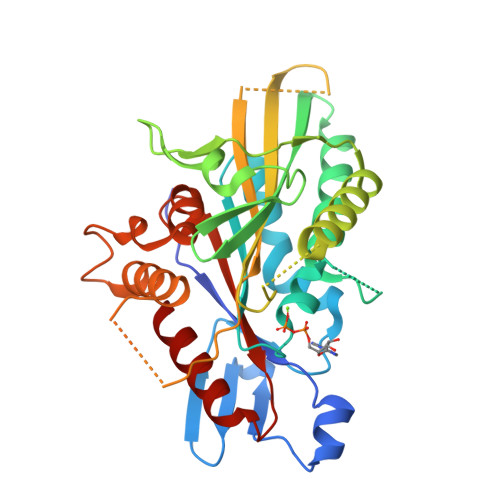
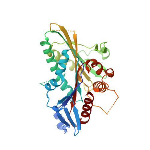
Structural Insights Into Human Kif7, a Kinesin Involved in Hedgehog Signalling.

Klejnot, M.Kozielski, F.

(2012) Acta Crystallogr D Biol Crystallogr 68: 154

  • DOI: https://doi.org/10.1107/S0907444911053042
  • Primary Citation of Related Structures:  
    2XT3, 4A14

  • PubMed Abstract: 

    Kif7, a member of the kinesin 4 superfamily, is implicated in a variety of diseases including Joubert, hydrolethalus and acrocallosal syndromes. It is also involved in primary cilium formation and the Hedgehog signalling pathway and may play a role in cancer. Its activity is crucial for embryonic development. Kif7 and Kif27, a closely related kinesin in the same subfamily, are orthologues of the Drosophila melanogaster kinesin-like protein Costal-2 (Cos2). In vertebrates, they work together to fulfil the role of the single Cos2 gene in Drosophila. Here, the high-resolution structure of the human Kif7 motor domain is reported and is compared with that of conventional kinesin, the founding member of the kinesin superfamily. These data are a first step towards structural characterization of a kinesin-4 family member and of this interesting molecular motor of medical significance.


  • Organizational Affiliation

    The Beatson Institute for Cancer Research, Garscube Estate, Switchback Road, Glasgow G61 1BD, Scotland. m.klejnot@beatson.gla.ac.uk


Macromolecules
Find similar proteins by:  (by identity cutoff)  |  3D Structure
Entity ID: 1
MoleculeChains Sequence LengthOrganismDetailsImage
KINESIN-LIKE PROTEIN KIF7344Homo sapiensMutation(s): 4 
UniProt & NIH Common Fund Data Resources
Find proteins for Q2M1P5 (Homo sapiens)
Explore Q2M1P5 
Go to UniProtKB:  Q2M1P5
PHAROS:  Q2M1P5
GTEx:  ENSG00000166813 
Entity Groups  
Sequence Clusters30% Identity50% Identity70% Identity90% Identity95% Identity100% Identity
UniProt GroupQ2M1P5
Sequence Annotations
Expand
  • Reference Sequence
Experimental Data & Validation

Experimental Data

  • Method: X-RAY DIFFRACTION
  • Resolution: 1.88 Å
  • R-Value Free:  0.229 (Depositor), 0.230 (DCC) 
  • R-Value Work:  0.179 (Depositor), 0.180 (DCC) 
  • R-Value Observed: 0.182 (Depositor) 
Space Group: P 21 21 21
Unit Cell:
Length ( Å )Angle ( ˚ )
a = 45.92α = 90
b = 79.79β = 90
c = 94.84γ = 90
Software Package:
Software NamePurpose
PHENIXrefinement
iMOSFLMdata reduction
SCALAdata scaling
MOLREPphasing

Structure Validation

View Full Validation Report


Ligand Structure Quality Assessment 

Created with Raphaël 2.3.0Worse 01 BetterLigand structure goodness of fit to experimental dataBest fitted ADPClick on this verticalbar to view details

Entry History 

Deposition Data

Revision History  (Full details and data files)

  • Version 1.0: 2011-12-14
    Type: Initial release
  • Version 1.1: 2012-01-25
    Changes: Other
  • Version 1.2: 2012-02-15
    Changes: Other
  • Version 1.3: 2023-12-20
    Changes: Data collection, Database references, Derived calculations, Other, Refinement description