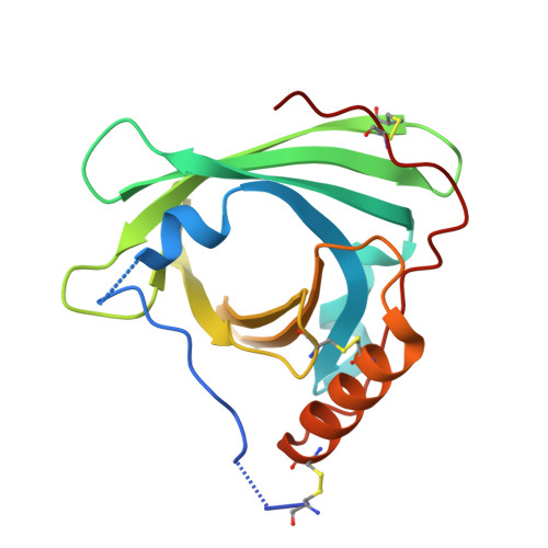
2YG2

Structure of apolioprotein M in complex with Sphingosine 1-Phosphate


Experimental Data Snapshot

  • Method: X-RAY DIFFRACTION
  • Resolution: 1.70 Å
  • R-Value Free: 0.221 
  • R-Value Work: 0.191 
  • R-Value Observed: 0.192 

Starting Model: experimental
View more details

wwPDB Validation   3D Report Full Report


Ligand Structure Quality Assessment 


This is version 1.4 of the entry. See complete history


Literature

Endothelium-Protective Sphingosine-1-Phosphate Provided by Hdl-Associated Apolipoprotein M.

Christoffersen, C.Obinata, H.Kumaraswamy, S.B.Galvani, S.Ahnstrom, J.Sevvana, M.Egerer-Sieber, C.Muller, Y.A.Hla, T.Nielsen, L.B.Dahlback, B.

(2011) Proc Natl Acad Sci U S A 108: 9613

  • DOI: https://doi.org/10.1073/pnas.1103187108
  • Primary Citation of Related Structures:  
    2YG2

  • PubMed Abstract: 

    Protection of the endothelium is provided by circulating sphingosine-1-phosphate (S1P), which maintains vascular integrity. We show that HDL-associated S1P is bound specifically to both human and murine apolipoprotein M (apoM). Thus, isolated human ApoM(+) HDL contained S1P, whereas ApoM(-) HDL did not. Moreover, HDL in Apom(-/-) mice contains no S1P, whereas HDL in transgenic mice overexpressing human apoM has an increased S1P content. The 1.7-Å structure of the S1P-human apoM complex reveals that S1P interacts specifically with an amphiphilic pocket in the lipocalin fold of apoM. Human ApoM(+) HDL induced S1P(1) receptor internalization, downstream MAPK and Akt activation, endothelial cell migration, and formation of endothelial adherens junctions, whereas apoM(-) HDL did not. Importantly, lack of S1P in the HDL fraction of Apom(-/-) mice decreased basal endothelial barrier function in lung tissue. Our results demonstrate that apoM, by delivering S1P to the S1P(1) receptor on endothelial cells, is a vasculoprotective constituent of HDL.


  • Organizational Affiliation

    Department of Clinical Biochemistry, Rigshospitalet, 2100 Copenhagen, Denmark.


Macromolecules
Find similar proteins by:  (by identity cutoff)  |  3D Structure
Entity ID: 1
MoleculeChains Sequence LengthOrganismDetailsImage
APOLIPOPROTEIN M
A, B
172Homo sapiensMutation(s): 0 
UniProt & NIH Common Fund Data Resources
Find proteins for O95445 (Homo sapiens)
Explore O95445 
Go to UniProtKB:  O95445
PHAROS:  O95445
GTEx:  ENSG00000204444 
Entity Groups  
Sequence Clusters30% Identity50% Identity70% Identity90% Identity95% Identity100% Identity
UniProt GroupO95445
Sequence Annotations
Expand
  • Reference Sequence
Small Molecules
Binding Affinity Annotations 
IDSourceBinding Affinity
FLC PDBBind:  2YG2 IC50: 900 (nM) from 1 assay(s)
Experimental Data & Validation

Experimental Data

  • Method: X-RAY DIFFRACTION
  • Resolution: 1.70 Å
  • R-Value Free: 0.221 
  • R-Value Work: 0.191 
  • R-Value Observed: 0.192 
  • Space Group: P 31 2 1
Unit Cell:
Length ( Å )Angle ( ˚ )
a = 68.28α = 90
b = 68.28β = 90
c = 135.58γ = 120
Software Package:
Software NamePurpose
REFMACrefinement
XDSdata reduction
XSCALEdata scaling
PHASERphasing

Structure Validation

View Full Validation Report


Ligand Structure Quality Assessment 


Entry History 

Revision History  (Full details and data files)

  • Version 1.0: 2011-06-01
    Type: Initial release
  • Version 1.1: 2011-06-16
    Changes: Version format compliance
  • Version 1.2: 2011-07-13
    Changes: Version format compliance
  • Version 1.3: 2023-12-20
    Changes: Data collection, Database references, Derived calculations, Other, Refinement description
  • Version 1.4: 2024-11-13
    Changes: Structure summary